Collection of materials relating to neuro-ophthalmology as part of the Neuro-Ophthalmology Virtual Education Library.
NOVEL: https://novel.utah.edu/
TO
- NOVEL966
Filters: Collection: "ehsl_novel_novel"
| Title | Creator | Description | Subject | ||
|---|---|---|---|---|---|
| 251 |
 |
Palatal Tremor | Shirley H. Wray, MD, PhD, FRCP | A PowerPoint slideshow describing the condition. | Bilateral Horizontal Gaze Palsy Hemorrhage; Bilateral Horizontal Gaze Palsy; Bilateral Lid Nystagmus; Bilateral Sixth Nerve Palsy; Brainstem Infarct; Degenerative Hypertrophy of the Inferior Olivary Nucleus; Facial Palsy; Facial Weakness; Lesion in the Guillain - Mollaret Triangle; Lid Nystagmus; O... |
| 252 |
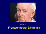 |
Frontotemporal Dementia | Shirley H. Wray, MD, PhD, FRCP | A PowerPoint slideshow describing the condition. | Acquired Ocular Motor Apraxia; Acquired Oculomotor Apraxia; CNS Degeneration; Complete paralysis of Voluntary Horizontal Saccades on Command to Look Left; Frontotemporal Dementia; Impaired Pursuit; Inability to Make a Refixation Saccade on Command to a Target Held on the Left; Normal Voluntary Hori... |
| 253 |
 |
Chiari-I Malformation | Shirley H. Wray, MD, PhD, FRCP | A PowerPoint slideshow describing the condition. | Ataxia; Chiari-1 Malformation; Downbeat Nystagmus; Dysmetria; Horizontal Saccadic Dysmetria; Lid Nystagmus; Oscillopsia; Primary Position Downbeat Nystagmus; Vertical Saccadic Dysmetria |
| 254 |
 |
Anterior Ischemic Optic Neuropathy (PowerPoint) | AAO/NANOS - American Academy of Ophthalmology / North American Neuro-Ophthalmology Society | The patient is a 62-year-old female who presented in August 1996 with visual loss OD that she first noted as loss of her superior field in May 1996. She felt that it had been static since, and perhaps was even a little better in the week before she was seen. There was no pain, even with ocular rotat... | Nonarteritic Ischemic Optic Neuropathy ; Anterior Ischemic Optic Neuropathy |
| 255 |
 |
Temporal Arteritis (PowerPoint) | AAO/NANOS - American Academy of Ophthalmology / North American Neuro-Ophthalmology Society | This 74-year-old asthmatic male had acute visual loss OS while watching the Super Bowl in 1994. He was seen the next day by a retina specialist, who noted that his optic disc was normal and referred the patient to a neuro-ophthalmologist, who evaluated him about 40 hours after his visual loss. He wa... | Temporal Arteritis |
| 256 |
 |
Optic Neuropathy with Sinus Disease (PowerPoint) | AAO/NANOS - American Academy of Ophthalmology / North American Neuro-Ophthalmology Society | The patient is a 66-year-old man with a history of ethanol abuse. He presented with 3 months of right-sided headache and a few days of progressive visual loss OD to hand motions only. When seen by the orbital service, he had nearly complete ophthalmoplegia and ptosis. Sinus biopsy showed fungus, whi... | Optic Neuropathy with Sinus Disease |
| 257 |
 |
Histiocytosis (PowerPoint) | AAO/NANOS - American Academy of Ophthalmology / North American Neuro-Ophthalmology Society | This 1-year-old child with familial erythrophagocytic lymphohistiocytosis was readmitted with a fever and was noted to have bilateral blindness. The spinal tap showed a protein of 148, with 178 WBC with 98% ""lymphocytes."" This image demonstrates the optic nerve infiltration. He was treated with ra... | Optic Nerve Histiocytosis; Histiocytosis |
| 258 |
 |
Orbital Tumors - Choroidal Folds From Orbital Mass (PowerPoint) | AAO/NANOS - American Academy of Ophthalmology / North American Neuro-Ophthalmology Society | This 30-year-old man had a retrobulbar intraconal mass OS. The CT scans showed a heterogeneous lobulated enhancing mass, 2.2 x 1.9 x 1.8 cm. The case beautifully exhibits chorodial folds. The ultrasound showed internal reflectivity. The patient refused surgery. | Choroidal Folds from Orbital Mass |
| 259 |
 |
Moyamoya Disease (PowerPoint) | AAO/NANOS - American Academy of Ophthalmology / North American Neuro-Ophthalmology Society | This 32-year-old woman was referred with a history of 4 days of loss of vision OD. She had a history of manic depressive illness and IV drug abuse; she had been HIV tested 4 weeks before and was negative. She said she last injected cocaine 5 days before being seen, the night before she awoke with th... | Saturday Night Retinopathy; Moyamoya Disease |
| 260 |
 |
Saturday Night Retinopathy (PowerPoint) | AAO/NANOS - American Academy of Ophthalmology / North American Neuro-Ophthalmology Society | This 32-year-old woman was referred with a history of 4 days of loss of vision OD. She had a history of manic depressive illness and IV drug abuse; she had been HIV tested 4 weeks before and was negative. She said she last injected cocaine 5 days before being seen, the night before she awoke with th... | Saturday Night Retinopathy |
| 261 |
 |
Moyamoya Syndrome (PowerPoint) | AAO/NANOS - American Academy of Ophthalmology / North American Neuro-Ophthalmology Society | A 9-year-old boy had recurrent ischemic episodes that had begun 2 years prior to evaluation. A significant right hemiparesis and a significant speech, learning, and memory disorder were present. His noncontrast axial view CT scan demonstrated multiple cerebral infarcts. Cerebral angiography revealed... | Moyamoya Disease; Moyamoya Syndrome |
| 262 |
 |
Periphlebitis in Optic Neuritis (PowerPoint) | AAO/NANOS - American Academy of Ophthalmology / North American Neuro-Ophthalmology Society | This 35-year-old otherwise-healthy woman developed typical optic neuritis OD with excellent recovery. She had no clinical evidence of multiple sclerosis at that time. She presented in August of 1991, at which time perivenous sheathing was seen in the retinal periphery OU. A limited workup was negati... | Periphlebitis in Optic Neuritis |
| 263 |
 |
Hereditary Optic Neuropathy (Leber's Hereditary Optic Neuropathy) | NANOS | Hereditary Optic Neuropathy - A hereditary optic neuropathy is caused by a genetic variant (or mutation) that causes dysfunction of the neurons (nerve cells) which form the optic nerve. The optic nerve sends information from the back of the eye to the vision center in the brain.The two most common t... | Hereditary Optic Neuropathy; Patient Brochure |
| 264 |
 |
Transient Vision Loss | NANOS | Vision loss that is temporary (transient) is a common problem and has many potential causes.Patients with temporary vision loss often do not have any abnormalities on their eye examination, especially once the vision has returned to normal. | Transient Vision Loss; Patient Brochure |
| 265 |
 |
NExT Introduction | Sachin Kedar, MD, Editor-in-Chief | Transcript of video introduction to the NExT curriculum collection. | NANOS Examination Techniques |
| 266 |
 |
Idiopathic Intracranial Hypertension | NANOS | Idiopathic intracranial hypertension (IIH), also called pseudotumor cerebri, is a condition in which there is high pressure in the fluid surrounding your brain, spinal cord, and optic nerves. This can cause headaches and problems with vision. | Idiopathic Intracranial Hypertension; Patient Brochure |
| 267 |
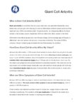 |
Giant Cell Arteritis | NANOS | Giant cell arteritis is a condition that can cause vision loss, new persistent headaches, scalp tenderness, and jaw pain with chewing. It is due to inflammation of blood vessels primarily of the head and neck. | Giant Cell Arteritis; Patient Brochure |
| 268 |
 |
Thyroid Eye Disease | NANOS | Thyroid eye disease, also called dysthyroid orbitopathy, is an autoimmune condition in whichyour body's immune system triggers inflammation in the eye socket (also called the orbit),affecting the muscles that move the eye and the fatty tissue behind the eye. | Thyroid Eye Disease; Thyroid Orbitopathy; Patient Brochure |
| 269 |
 |
Metastatic Glioblastoma to Intracranial Optic Nerves, Optic Chiasm and Optic Tracts | Bashaer Aldhahwani, MD; Mariam S. Vilá-Delgado, MD | The patient with pathology confirmed glioblastoma after resectioning the superior frontal lobe tumor followed by 6 weeks of radiation therapy with concurrent temozolomide. The patient started bevacizumab to treat steroid-refractory vasogenic cerebral edema/radiation necrosis. 8 months after radiatio... | Metastatic Glioblastoma; Infiltrative Chiasmal Lesion |
| 270 |
 |
Vascular Headache Disorders | Salman Ali Mahmood, MD; Sean Gratton, MD | This is a PowerPoint with embedded audio presenting a detailed summary of the evaluation and management of an important cause of secondary headaches: vascular headache disorders. Topics covered include intracranial hemorrhage, arterial dissection, giant cell arteritis, cerebral venous sinus thrombos... | Vascular Headache Disorders; Epidural Hematoma; Subdural Hematoma; Subarachnoid Hemorrhage; Intracranial Hemorrhage; Arterial Dissection; Giant Cell Arteritis; Cerebral Venous Sinus Thrombosis; Pituitary Apoplexy |
| 271 |
 |
Recurrent Painful Ophthalmoplegic Neuropathy | Jay Chopra, BS; Devin D. Mackay, MD | Overview of recurrent painful ophthalmoplegic neuropathy with an illustrative case example and discussion of clinical presentation, possible mechanisms, and treatment. | Recurrent Painful Ophthalmoplegic Neuropathy; RPON; Ophthalmoplegic Migraine; Ophthalmoparesis; Painful Cranial Nerve Palsy |
| 272 |
 |
Tacrolimus Optic Neuropathy | Hailey Mair, BS; Padmaja Sudhakar, MD | This PowerPoint slide deck will describe tacrolimus optic neuropathy which is a very rare form of optic neuropathy but remains a potential issue in many patients that receive this drug. | Optic Neuropathy; Tacrolimus; Tacrolimus Associated Visual Loss |
| 273 |
 |
Natural Language Processing in Neuro-Ophthalmology | Areeba Abid, BS; Sachin Kedar, MD | This video provides an overview of natural language processing (NLP) techniques, applications, and limitations in neuro-ophthalmology. NLP, a branch of artificial intelligence, enables computers to understand and analyze human language. This video discusses three types of NLP techniques: sentiment a... | Artificial Intelligence; Machine Learning; Natural Language Processing; Word Prediction; Word Embeddings; Sentiment Analysis |
| 274 |
 |
Lessons From Bench Bedside | Shirley H. Wray, MD, PhD, FRCP | See also: http://content.lib.utah.edu/cdm/ref/collection/ehsl-shw/id/69, http://content.lib.utah.edu/cdm/ref/collection/ehsl-shw/id/282, http://content.lib.utah.edu/cdm/ref/collection/ehsl-shw/id/94, and http://content.lib.utah.edu/cdm/ref/collection/ehsl-shw/id/103 | Bilateral Internuclear Ophthalmoplegia; Pendular Horizontal Oscillations; Lid Nystagmus; Upbeat Nystagmus; Botulinum Toxin Therapy; Multiple Sclerosis; Horizontal Pendular Nystagmus; Gaze Evoked Upbeat Nystagmus; Abducting Nystagmus; Normal Convergence; Gaze Evoked Downbeat Nystagmus; Sac... |
| 275 |
 |
Optochiasmal Tuberculoma | Jeanie Paik, MD; Rudrani Banik, MD | PowerPoint of case of chiasmal tuberculoma causing bitemporal defect in patient with tuberculosis on RIPE treatment; case history, differential diagnosis and treatment discussed. | Chiasmal Disorder; Chiasmal Tuberculoma; Bitemporal Visual Field Defect; Ethambutol Optic Neuropathy |
