Collection of materials relating to neuro-ophthalmology as part of the Neuro-Ophthalmology Virtual Education Library.
NOVEL: https://novel.utah.edu/
TO
- NOVEL966
Filters: Collection: "ehsl_novel_novel"
| Title | Creator | Description | Subject | ||
|---|---|---|---|---|---|
| 251 |
 |
Olfactory System: Neuroanatomy Video Lab - Brain Dissections | Suzanne S. Stensaas, PhD | Beginning with the location of the sensory cells within the skull the axons are traced into the cranial cavity. Demonstration of the olfactory bulb, olfactory tract and it termination in the forebrain and temporal lobe are indicated. Trauma and meningiomas can produce loss of small (anosmia). Degene... | Olfactory System; Olfactory Bulb; Anosmia; Brain; Dissection |
| 252 |
 |
The Meninges: Neuroanatomy Video Lab - Brain Dissections | Suzanne S. Stensaas, PhD | The epidural, subdural and subarachnoid spaces are demonstrated and discussed with respect to trauma and disease. The relationship of the brainstem and cerebellum to the tentorium demonstrates the vulnerability of the brain stem to increased supratentorial pressure and herniation. Arachnoid granulat... | Meninges; Brain; Dissection |
| 253 |
 |
Orientation: The Planes of the Brain: Neuroanatomy Video Lab - Brain Dissections | Suzanne S. Stensaas, PhD | Terms such as anterior, posterior, inferior and superior are introduced with respect to the hemispheres as well as the brain stem. Terms such as rostral and caudal or dorsal and ventral can mean different things in different areas. Sections in three planes (frontal, axial, and sagittal) are demonstr... | Frontal; Axial; Sagittal; Brain; Dissection |
| 254 |
 |
The Visual Pathway: Neuroanatomy Video Lab - Brain Dissections | Suzanne S. Stensaas, PhD | A brief review of the anatomy of the eye and the photic stimulation of the receptors is followed by a gross exploration of the visual pathway from the optic nerve, chiasm, and tract to the thalamus stressing how the left part of the visual world reaches the right hemisphere. Visual fields are relate... | Visual Pathway; Brain; Dissections |
| 255 |
 |
Ischemic Ocular Syndrome | Kathleen B. Digre, MD; James J. Corbett, MD | Slideshow describing condition. | Ischemic Ocular Syndrome |
| 256 |
 |
The Normal Unfixed Brain: Neuroanatomy Video Lab - Brain Dissections | Suzanne S. Stensaas, PhD | The consistency and vulnerability of the brain is demonstrated along with the clear and glistening pia and arachnoid and the tough dura. The cushioning function of the CSF is stressed and the features are pointed out on the ventral surface. The uncus and temporal lobes are normal with arteries free ... | Brain; Dissections |
| 257 |
 |
Multifocal Choroiditis | Gregory P. Van Stavern, MD | Multi-focal choroiditis is usually a bilateral choroidopathy seen more frequently in women associated with punched out appearing lesions occasionally with pigment around the edges. Image provides example. | Multi-Focal Choroiditis Panuveitis |
| 258 |
 |
Retinitis Pigmentosa | Gregory P. Van Stavern, MD | Retinitis pigmentosa is a retinal/choroidal degeneration caused by various genetic defects. The term retinitis pigmentosa is really a misnomer since it is not inflammation (retinitis) and it is not a disease of the pigmentary system (pigmentosa). | Retinitis Pigmentosa |
| 259 |
 |
Serpiginous Choroidopathy | Gregory P. Van Stavern, MD | Serpiginous choroidopathy (also known as Geographic choroidopathy) usually affects the choroid, the choriocapillaris and the retinal pigment epithelium in both eyes. | Serpiginous Choroidopathy |
| 260 |
 |
Birdshot | Gregory P. Van Stavern, MD | Birdshot Retinochoroidopathy is a posterior uveitis seen in women 30-60 years of age who present with floaters, changes in color vision, and difficulty with night vision. | Birdshot Choroidopathy |
| 261 |
 |
Pars Planitis | Gregory P. Van Stavern, MD | Pars planitis is an inflammatory condition seen in children and young adults. It is associated with inflammation of the pars plana--at the far periphery of the retina. | Pars Planitis |
| 262 |
 |
Vogt Koyanagi-Harada (VKH) Syndrome | Gregory P. Van Stavern, MD | Vogt-Koyanagi disease causes bilateral uveitis, along with alopecia, vitiligo, and hearing loss. | Vogt Koyanagi-Harada Syndrome (VKH) |
| 263 |
 |
Stargardt's Disease | Gregory P. Van Stavern, MD | Stargardt's disease is an inherited maculopathy which frequently presents with a loss of central vision. | Stargardt's Disease |
| 264 |
 |
Pontine_Infarction | Shirley H. Wray, MD, PhD, FRCP | The patient is a 62 year old right handed man, status post myocardial infarction in 1989 and on Coumadin. In 1993 he presented with a history of three separate TIAs 1.Instantaneous perioral tingling and/or numbness lasting less than 1 minute. 2.Episodic numbness of the right hand and foot lasting le... | Unilateral Internuclear Ophthalmoplegia; Unilateral Horizontal Gaze Palsy; Upbeat Nystagmus on Upgaze; Convergence Normal; Fisher's One-and-a-Half Syndrome; Pontine Infarct; Unilateral Horizontal Gaze Palsy Infarct; Abducting Nystagmus |
| 265 |
 |
Best's Vittelform Maculopathy | Gregory P. Van Stavern, MD | This 14 year old presented with decreased vision, headaches and central scotomas. She was found to have bilateral papilledema related to IIH and also Best's vitilliform maculopathy. The maculas are commonly described as having a "fried egg" sunny side up appearance. | Best Macular Dystrophy |
| 266 |
 |
Parinaud Syndrome | Raed Behbehani, MD | Parinaud syndrome, as called dorsal midbrain syndrome, is due to dorsal midbrain lesions from compression (e.g., a tumor), demyelination, or ischemia. The syndrome is characterized by limitation of upward gaze, convergence retraction nystagmus, light near dissociation, and lid retraction (Collier's ... | Dorsal Mibrain Syndrome; Parinaud's Syndrome |
| 267 |
 |
Square Wave Jerks with Contrapulsion | Raed Behbehani, MD | A patient with history of brain stem stroke 2 months ago (right hemifacial anesthesia , left sided weakness and bulbar symptoms dysphagia) comes complaining of oscillipsia , binocular vertical diplopia). On exam he had a vertical tropia of 3-4 PD (Skew deviation), dissociated nystagmus , and saccadi... | Square Wave Jerks; Contrapulsion |
| 268 |
 |
Oculopalatal Tremor | Raed Behbehani, MD | This is a usually vertical, pendular nystagmus associated with synchronous rhythmic movement of the palate, developing months after a severe brain stem stroke. The stroke involves the dentato-rubro-olivary tract (Mollaret's triangle). MRI can show hypertrophy of the inferior olivary nucleus in the m... | Oculopalatal Tremor |
| 269 |
 |
Constructional Apraxia | Shirley H. Wray, MD, PhD, FRCP | The patient is a 72 year old right handed woman who presented in November 1995 with the sudden onset of impaired coordination of visual and motor skills following an inner right ear infection. One of her problems was difficulty sitting on a chair as she tended to place her body incorrectly. By late ... | Dressing Apraxia; Apraxia of the Left Hand; Constructional Apraxia; Right Parietal Lobe; Progressive Lobar Atrophy; Degenerative CNS Disease; Apraxia |
| 270 |
 |
Downbeat Nystagmus PAN | Shirley H. Wray, MD, PhD, FRCP | This patient carries a diagnosis of multiple sclerosis. See also: http://content.lib.utah.edu/cdm/ref/collection/ehsl-shw/id/317 | Downbeat Nystagmus; Periodic Alternating Nystagmus; Multiple Sclerosis; Primary Position Downbeat Nystagmus |
| 271 |
 |
Brain Control of Horizontal Saccadic Eye Movements (Guest Lecture) | Shirley H. Wray, MD, PhD, FRCP | In 1995 I published this case alongside eleven personal cases, three with the Kearns-Sayer Syndrome (KSS) and five with Progressive External Opthalmoplegia (PEO). Am J of Neuroradiol:16 (5);1167-1173. The patient was under the care of Dr. Raymond Adams from age 13 years. In 1991, at age 40 years, I ... | Bilateral Ptosis; Facial Weakness; Complete External Ophthalmoplegia; Bilateral Progressive External Ophthalmoplegia (PEO); Mitochondrial Myopathy; PEO plus Deafness; Cerebellar Degeneration with Ataxia; Chronic Progressive External Opthalmoplegia |
| 272 |
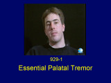 |
Essential Palatal Tremor | Shirley H. Wray, MD, PhD, FRCP | The patient is a 25 year old meteorologist from Tennessee who came to Boston in the summer of 1992 to vacation with his family on the Cape. His illness started with flu-like symptoms, low grade fever from 99 to 100F, sweating and episodes of light headedness associated with occasional nausea, indige... | Essential Palatal Tremor (Myoclonus); Brainstem Encephalitis |
| 273 |
 |
Palatal Tremor | Shirley H. Wray, MD, PhD, FRCP | See also: http://content.lib.utah.edu/cdm/ref/collection/ehsl-shw/id/68, http://content.lib.utah.edu/cdm/ref/collection/ehsl-shw/id/247, http://content.lib.utah.edu/cdm/ref/collection/ehsl-shw/id/111, http://content.lib.utah.edu/cdm/ref/collection/ehsl-shw/id/312, http://content.lib.utah.edu/cdm/ref... | Palatal Tremor (Myoclonus); Pendular Vertical Oscillations; Unilateral Horizontal Gaze Palsy; Facial Palsy; Pontine Infarct; Degenerative Hypertrophy of the Inferior Olivary Nucleus; Lesion in the Guillain-Mollaret Triangle; Oculopalatal Myoclonus; Oculopalatal Tremor Lid Nystagmus; Bilate... |
| 274 |
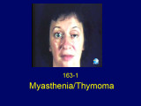 |
Myasthenia Thymoma | Shirley H. Wray, MD, PhD, FRCP | The patient is a 46 year old woman who presented in July 1977 with horizontal double vision lasting two weeks. Three weeks later the left upper eyelid started to droop and by the end of the day the eye was closed. She had no ptosis of the right eye and no generalized fatigue. She consulted an intern... | Unilateral Ptosis; Unilateral Lid Retraction; Myasthenic Lid Twitch; External Ophthalmoplegia; Ocular Myasthenia Gravis; Tensilon Test; Thymolipoma; Generalized Myasthenia Gravis; Unilateral Myasthenia Gravis; Myasthenic Ptosis; Lid Retraction; Lid Twitch |
| 275 |
 |
Multiple Sclerosis | Shirley H. Wray, MD, PhD, FRCP | The patient is a 56 year old woman who presented in 1982, at the age of 48, with a one week history of painless loss of vision in the left eye. Past History: Negative for a previous attack of optic neuritis or transient neurological symptoms. Family History: Negative for CNS disease Neuro-ophthalmol... | Upbeat Nystagmus; Lid Nystagmus; Square Wave Jerks; Jerk Oscillations; Rotary Nystagmus; Saccadic Pursuit; Saccadic Dysmetria; Multiple Sclerosis; Bilateral Lid Nystagmus; Primary Position Upbeat Nystagmus; Torsional Nystagmus; Horizontal Saccadic Dysmetria |