AAO-NANOS Neuro-Ophthalmology Clinical Collection: Derived from the AAO-NANOS Clinical Neuro-Ophthalmology collection produced on CD. The images are of selected cases from the NANOS teaching slide exchange, and the CD was produced under the direction of Larry Frohman, MD and Andrew Lee, MD.
The American Academy of Ophthalmology (AAO); The North American Neuro-Ophthalmology Association (NANOS).
NOVEL: https://novel.utah.edu/
TO
Filters: Collection: ehsl_novel_aao_nanos
| Title | Description | Subject | ||
|---|---|---|---|---|
| 226 |
 |
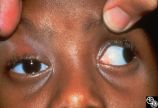
Motility Disturbances | Cyclical oculomotor paresis may occur in patients as an intermittent phenomenon, with a paretic phase and diplopia and intervals that are nonparetic. The history and examination are classic for the disorder. Pair with Images 95_18 and 95_20. | Oculomotor Palsy (Cyclical Oculomotor Palsy); Cyclical |
| 227 |
 |
Motility Disturbances | Cyclical oculomotor paresis may occur in patients as an intermittent phenomenon, with a paretic phase and diplopia and intervals that are nonparetic. The history and examination are classic for the disorder. Pair with Images 95_18 and 95_19. | Oculomotor Palsy (Cyclical Oculomotor Palsy); Cyclical |
| 228 |
 |
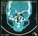
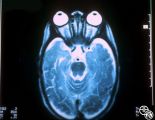
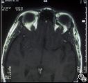
Neuro-Ophthalmic Imaging-MRI | Aneurisms may result in neuro-ophthalmologic sign and symptoms by direct compression of the afferent or efferent systems or by the secondary effects of hemorrhage. Basilar aneurisms may result in ocular motor deficits such as a unilateral or bilateral third nerve palsy. | Aneurysm |
| 229 |
 |
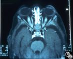
Neuro-Ophthalmic Imaging-MRI | Aneurisms may result in neuro-ophthalmologic sign and symptoms by direct compression of the afferent or efferent systems or by the secondary effects of hemorrhage. Basilar aneurisms may result in ocular motor deficits such as a unilateral or bilateral third nerve palsy. | Aneurysm |
| 230 |
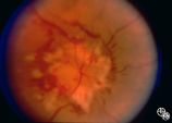 |
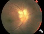
Systemic Disorders With Optic Nerve and Retinal Findings | Neoplasms may result in an optic neuropathy by direct metastatic involvement. In this patient, a lung adenocarcinoma was metastatic to the optic nerve.This is a fundus photo. | Metastatic Carcinoma; Metastasis; Optic Nerve |
| 231 |
 |
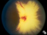
Isolated Congenital Optic Disc Anomalies | This patient has optic disc drusen and evidence of a superimposed optic neuropathy, including loss of visual field, an ipsilateral afferent pupillary defect, and optic atrophy. Although optic disc drusen typically causes visual field loss without visual acuity loss superimposed, ischemic optic neuro... | Optic Disc Drusen; Optic Nerve Drusen; Optic Neuropathy |
| 232 |
 |
Isolated Congenital Optic Disc Anomalies | Normally, there is no visible myelination of the nerve fiber layer in the retina. Occasionally, visible myelination occurs that takes on a characteristic white, arcuate, feathery appearance that follows the contour of the nerve fiber layer. Disease/Diagnosis: Myelinated Nerve Fiber. | Myelinated Nerve Fiber Layer |
| 233 |
 |
Acquired Disc Changes | Although optociliary shunt vessels are venous collaterals that form in response to chronic venous obstruction, they may occur in patients with chronic open-angle glaucoma. | Shunt Vessels (Glaucoma) |
| 234 |
 |
Acquired Disc Changes | Optociliary shunt vessels are venous collaterals that form in response to chronic venous obstruction. They may occur in patients following central retinal vein occlusion. | Shunt Vessels (CRVO) |
| 235 |
 |
Acquired Disc Changes | Although optociliary shunt vessels are venous collaterals that typically form in response to chronic venous obstruction, they may occur on a congenital basis as seen here. | Shunt Vessels (Congenital) |
| 236 |
 |
Acquired Disc Changes | Optociliary shunt vessels are venous collaterals that form after chronic venous obstruction. The presence of optic atrophy, progressive visual loss, and optociliary shunt vessels may indicate a compressive optic nerve lesion such as meningioma. | Shunt Vessels (Meningioma) |
| 237 |
 |
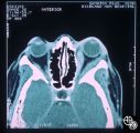
Neuro-Ophthalmic Imaging-CT Scan | This is a patient with trauma leading to enucleation, with swelling years later over the implant. This is a presumed chronic abscess between orbit and dura. | Orbital Abscess |
| 238 |
 |
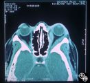
Neuro-Ophthalmic Imaging-CT Scan | Idiopathic orbital pseudotumor is an inflammatory disorder that may effect any part of the ocular anatomy. The site of inflammation determines the nomenclature. For example, involvement of the sclera is referred to as scleritis. And involvement of one or more of the extraocular muscles is referred t... | Myositis; Orbital Inflammation |
| 239 |
 |
Neuro-Ophthalmic Imaging-CT Scan | Idiopathic orbital pseudotumor is an inflammatory disorder that may effect any part of the ocular anatomy. The site of inflammation determines the nomenclature. For example, involvement of the sclera is referred to as scleritis. And involvement of one or more of the extraocular muscles is referred t... | Myositis |
| 240 |
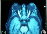 |
Systemic Disorders With Optic Nerve and Retinal Findings | This 1-year-old child with familial erythrophagocytic lymphohistiocytosis was readmitted with a fever and was noted to have bilateral blindness. The spinal tap showed a protein of 148, with 178 WBC with 98% ""lymphocytes."" This MRI image demonstrates the optic nerve infiltration. He was treated wit... | Optic Nerve Histiocytosis; MRI Orbit; Optic Nerves |
| 241 |
 |
Systemic Disorders With Optic Nerve and Retinal Findings | This 1-year-old child with familial erythrophagocytic lymphohistiocytosis was readmitted with a fever and was noted to have bilateral blindness. The spinal tap showed a protein of 148, with 178 WBC with 98% ""lymphocytes."" This MRI image demonstrates the optic nerve infiltration. He was treated wit... | Optic Nerve Histiocytosis; MRI Orbit; Optic Nerves |
| 242 |
 |
Systemic Disorders With Optic Nerve and Retinal Findings | This 1-year-old child with familial erythrophagocytic lymphohistiocytosis was readmitted with a fever and was noted to have bilateral blindness. The spinal tap showed a protein of 148, with 178 WBC with 98% ""lymphocytes."" This MRI image demonstrates the optic nerve infiltration. He was treated wit... | Optic Nerve Histiocytosis; MRI Orbit; Optic Nerves |
| 243 |
 |
Systemic Disorders With Optic Nerve and Retinal Findings | This 25-year-old man presented to the eye service with a history of 3 days of decreased vision OD. His past medical history was unremarkable. His examination showed acuities of 20/25 OU, with intact color plates, a 0.3 log unit of RAPD OD, and an inferior arcuate scotoma. The photos (Images 95_42, 9... | Toxoplasmosis |
| 244 |
 |
Neuro-Ophthalmic Vascular Disease | This 27-year-old woman had no past ocular history and presented with 3 weeks of redness OS that has been treated by the referring doctor as allergic conjunctivitis. She was referred for evaluation when she developed binocular diplopia. Her past medical history included phlebitis and one miscarriage ... | Superior Ophthalmic Vein Thrombosis |
| 245 |
 |
Neuro-Ophthalmic Vascular Disease | This 27-year-old woman had no past ocular history and presented with 3 weeks of redness OS that has been treated by the referring doctor as allergic conjunctivitis. She was referred for evaluation when she developed binocular diplopia. Her past medical history included phlebitis and one miscarriage ... | Superior Ophthalmic Vein Thrombosis |
| 246 |
 |
Neuro-Ophthalmic Vascular Disease | This 27-year-old woman had no past ocular history and presented with 3 weeks of redness OS that has been treated by the referring doctor as allergic conjunctivitis. She was referred for evaluation when she developed binocular diplopia. Her past medical history included phlebitis and one miscarriage ... | Superior Ophthalmic Vein Thrombosis |
| 247 |
 |
Systemic Disorders With Optic Nerve and Retinal Findings | This 74-year-old asthmatic male had acute visual loss OS while watching the Super Bowl in 1994. He was seen the next day by a retina specialist, who noted that his optic disc was normal and referred the patient to a neuro-ophthalmologist, who evaluated him about 40 hours after his visual loss. He wa... | Temporal Arteritis |
| 248 |
 |
Systemic Disorders With Optic Nerve and Retinal Findings | This 74-year-old asthmatic male had acute visual loss OS while watching the Super Bowl in 1994. He was seen the next day by a retina specialist, who noted that his optic disc was normal and referred the patient to a neuro-ophthalmologist, who evaluated him about 40 hours after his visual loss. He wa... | Temporal Arteritis |
| 249 |
 |
Systemic Disorders With Optic Nerve and Retinal Findings | This 74-year-old asthmatic male had acute visual loss OS while watching the Super Bowl in 1994. He was seen the next day by a retina specialist, who noted that his optic disc was normal and referred the patient to a neuro-ophthalmologist, who evaluated him about 40 hours after his visual loss. He wa... | Temporal Arteritis |
| 250 |
 |
Systemic Disorders With Optic Nerve and Retinal Findings | This 74-year-old asthmatic male had acute visual loss OS while watching the Super Bowl in 1994. He was seen the next day by a retina specialist, who noted that his optic disc was normal and referred the patient to a neuro-ophthalmologist, who evaluated him about 40 hours after his visual loss. He wa... | Temporal Arteritis |