Best known for his world-renowned neuro-ophthalmology unit based at the University of California, San Francisco, William Hoyt, MD collected here more than 850 of his best images covering a wide range of disorders.
William F. Hoyt, MD, Professor Emeritus of Ophthalmology, Neurology and Neurosurgery, Department of Ophthalmology, University of California, San Francisco.
NOVEL: https://novel.utah.edu/
TO
Filters: Collection: "ehsl_novel_wfh"
| Title | Description | Type | ||
|---|---|---|---|---|
| 226 |
 |
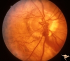
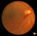
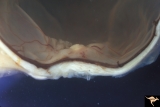
Segmental Atrophy - Hemianopic (Band) Atrophy | Segmental Atrophy - Band atrophy with temporal hemianopia. 1983. Anatomy: Optic disc. Pathology: Atrophy of the chiasm or left optic tract. Disease/Diagnosis: Segmental band atrophy. Clinical: Right temporal field defect. | Image |
| 227 |
 |
G208 Traumatic AION | Traumatic vitreopapillary evulsion (traumatic AION). Traumatic AION from evulsion of the vitreopapillary adhesion. Leakage on disc surface where vitreous was adherent. Pair with G2_9b. Anatomy: Optic disc. Pathology: AION. Disease/ Diagnosis: Traumatic AION. | Image |
| 228 |
 |
G209 Traumatic AION | Traumatic vitreopapillary evulsion (traumatic AION). Fluorescein angiogram shows petal shaped avascular zones on the surface of the disc. Pair with G2_8a. Anatomy: Optic disc. Pathology: AION. Disease/ Diagnosis: Traumatic AION. Imaging: Flourescein angiogram. | Image |
| 229 |
 |
C403 Luetic Papillopathy (Gumma of the Optic Disc) | 40 year old man with AIDS and neurosyphillis with severe visual field defect. The disc is pale and swollen and its arteries are strikingly narrowed (syphillitic vasculitis). Anatomy: Optic disc. Pathology: Axoplasmic stasis due to syphillitic infection. Disease/ Diagnosis: Luetic papillopathy (Syphy... | Image |
| 230 |
 |
ID05a Post Papilledema Optic Atrophy from Pseudotumor Cerebri | Left eye, October 1999, Post papilledema optic atrophy from pseudotumor cerebri. Note optociliary veins in both discs. Gliosis and partial pallor following long standing papilledema and intracranial pressure. Anatomy: Optic disc. Pathology: Post papilledema atrophy and gliosis from long standing el... | Image |
| 231 |
 |
ID05b Post Papilledema Optic Atrophy from Pseudotumor Cerebri | Right eye, October 1999, Post papilledema optic atrophy from pseudotumor cerebri. Note optociliary veins in both discs. Gliosis and partial pallor following long standing papilledema and intracranial pressure. Anatomy: Optic disc. Pathology: Post papilledema atrophy and gliosis from long standing el... | Image |
| 232 |
 |
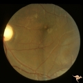
Venous Anomalies - Exit Anomalies | Choriovaginal vein. Inferior portion of disc at 6:00. Wide angle view of same patient as V_41. Anatomy: Optic disc. Pathology: Congenital anomaly of choroidal venous drainage. Disease/Diagnosis: Choriovaginal vein. Clinical: Asymptomatic. | Image |
| 233 |
 |
Venous Anomalies - Exit Anomalies | Choriovaginal vein. Inferior portion of disc at 5:00. Same patient as V_42. Anatomy: Optic disc. Pathology: Congenital anomaly of choroidal venous drainage. Disease/Diagnosis: Choriovaginal vein. Clinical: Asymptomatic. | Image |
| 234 |
 |
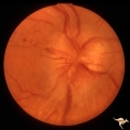
Venous Anomalies - Exit Anomalies | Anomalous exits of retinal veins at 5:00 . Anatomy: Optic disc. Pathology: Congenital anomaly, exit anomaly. Disease/Diagnosis: Exit anomaly, edge veins. Clinical: Asymptomatic. | Image |
| 235 |
 |
Diffuse Atrophy | Nerve fiber appearance about 6 weeks after indirect injury to optic nerve. Note near total absence of nerve fiber reflexes. Photo shows remaining streaks of inferior arcuate nerve fiber membranes dissolving into nothing. 1972. Anatomy: Optic disc. Pathology: Optic nerve injury. Disease/Diagnosis: Op... | Image |
| 236 |
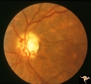 |
IA03 Atrophy with Optociliary Veins | 1974, left eye, perioptic nerve sheath meningioma, blind eye. Optociliary bypass veins in the nasal disc tissue. Anatomy: Optic disc. Pathology: Optociliary vein. DIsease/ Diagnosis: Perioptic nerve sheath meningioma. Clinical: Blind eye. | Image |
| 237 |
 |
Vascular Disc Anomalies - Retinal Arteriovenous Malformations | Retinal arteriovenous malformations. Composite photograph. Notice there are at least two complete loops, nasally and infratemporally. Notice the signs of early involution centrally (white). Anatomy: Optic disc. Pathology: Retinal arteriovenous malformation. Disease/Diagnosis: Retinal arteriovenous m... | Image |
| 238 |
 |
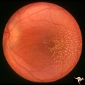
Vascular Disc Anomalies - Retinal Arteriovenous Malformations | Retinal arteriovenous malformations showing a complete arteriovenous loop nasally. 19 year old female. Composite photograph. Anatomy: Optic disc. Pathology: Retinal arteriovenous malformation. Disease/Diagnosis: Retinal arteriovenous malformation. Clinical: Asymptomatic. | Image |
| 239 |
 |
IB108 Post Ischemic (AION) Cupless Atrophy | March 1978, right eye, patient had inferior field defect, top half of optic nerve is atrophic, hemiatrophy, blood vessels are focally narrow, bottom half of disc is normal. Anatomy: Optic disc. Pathology: Post ischemic (AION) cupless atrophy. Disease/ Diagnosis: Post ischemic (AION) cupless atrophy.... | Image |
| 240 |
 |
Segmental Atrophy - Temporal | Segmental Atrophy - Temporal - Nutritional Amblyopia - Nerve fiber layer hemorrhage. Pair with IIA2_04b. 1970. Anatomy: Optic disc. Pathology: Optic atrophy. Disease/Diagnosis: Toxic optic atrophy from alcohol. Clinincal: Central visual loss. | Image |
| 241 |
 |
Segmental Atrophy - Temporal | Temporal - Nutritional Amblyopia - Nerve fiber layer hemorrhage. Pair with IIA2_04a. 1970. Anatomy: Optic disc. Pathology: Optic atrophy. Disease/Diagnosis: Toxic optic atrophy from alcohol. Clinical: Central visual loss. | Image |
| 242 |
 |
C201 Papillitis with Macular Star, Cat Scratch Disease | Proven Bartonella neuroretinitis. Notice the deposit of exudates of Henle's layer making an almost complete macular star. Anatomy: Optic disc; Retina. Pathology: Neuroretinitis; Axoplasmic stasis due to inflammation. Disease/ Diagnosis: Neuroretinitis due to Bartonella Henslae. Clinical: Visual blur... | Image |
| 243 |
 |
B110 Disc Swelling, Ischemic Papillopathies, AION | Pallid ischemic swelling with intraretinal exudates near the macula and a ""cotton wool"" infarct below the disc. 38 year old man. Diabetic. 20/60 vision. Altitudinal visual field defect. Anatomy: Optic disc. Pathology: Axoplasmic stasis due to ischemia. Disease/ Diagnosis: AION. Clinical: Diabetic ... | Image |
| 244 |
 |
B113 Disc Swelling, Ischemic Papillopathies, AION | 57 year old woman with AION. Anatomy: Optic disc. Pathology: Axoplasmic stasis due to ischemia. Disease/ Diagnosis: AION. Clinical: Visual loss. | Image |
| 245 |
 |
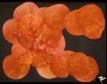
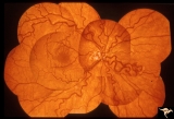
F2b10 Malignant Optic Nerve Glioma, Gross Pathologic Specimen | Pathologic specimen of optic nerve glioma shown in slide F2b_09. White material on top of swollen disc is myelin. Reference: Hoyt WF, Meshel LG, Lessell S, Schatz NJ, Suckling RD. Malignant optic glioma of adulthood. Brain. 1973;96(1):121-32. Anatomy: Optic disc. Pathology: Optic nerve glioma. Disea... | Image |
| 246 |
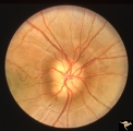 |
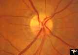
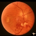
Buried Drusen | 7 year old boy with pseudo papilledema from buried drusen. Note the lumpy contour of the disc margin. Also note the surrounding ring-like light reflex that is optically perfect and indicates absence of edema spreading onto the surrounding retina. Anatomy: Optic disc. Pathology: Drusen of the optic d... | Image |
| 247 |
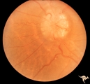 |
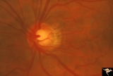
Chronic Papilledema with Pseudo Drusen | Right eye. Meningioma. Pseudo drusen from chronic papilledema. Woman. Anatomy: Optic disc Pathology: Papilledema Disease/Diagnosis: Chronic papilledema with pseudo drusen | Image |
| 248 |
 |
Chronic Papilledema with Pseudo Drusen | Left eye. Meningioma. Pseudo drusen from chronic papilledema. The patient's meningioma had blinded her left eye and caused chronic elevated intracranial pressure. Woman. Anatomy: Optic disc Pathology: Papilledema Disease/Diagnosis: Chronic papilledema with pseudo drusen | Image |
| 249 |
 |
F107 Metastatic Breast Cancer to the Disc | Metastatic breast cancer to the disc. Notice mass on inferior portion of disc. Also notice tangled capillary dilation within the mass indicating infiltration. This disc tumor was radiated. It disappeared leaving a pale flat atrophic nerve. The patient died. Histologic study of the eye revealed metas... | Image |
| 250 |
 |
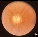
Buried Drusen with Choroidal Retinal Scar | Right eye: Buried drusen; probable complication of peripapillary hemorrhage at 7:00. Anatomy: Optic disc. Pathology: Drusen of the optic disc. Disease/Diagnosis: Drusen of the optic disc. Clinical notes: Enlarged blind spot. | Image |