Best known for his world-renowned neuro-ophthalmology unit based at the University of California, San Francisco, William Hoyt, MD collected here more than 850 of his best images covering a wide range of disorders.
William F. Hoyt, MD, Professor Emeritus of Ophthalmology, Neurology and Neurosurgery, Department of Ophthalmology, University of California, San Francisco.
NOVEL: https://novel.utah.edu/
TO
Filters: Collection: "ehsl_novel_wfh"
| Title | Description | Type | ||
|---|---|---|---|---|
| 226 |
 |
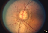
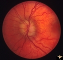
Bilateral Severe Hemorrhagic Papilledema | Right eye. Bilateral hyperacute papilledema with rapid blindness associated with dural sinus occlusion. Both eyes were nearly blind. Young man. Anatomy: Optic disc. Pathology: Papilledema. Disease/Diagnosis: Bilateral hyperacute papilledema | Image |
| 227 |
 |
Bilateral Severe Hemorrhagic Papilledema | Left eye. Two months later, resolving Bilateral Severe Hemorrhagic Papilledema. Same eye as P_32b | Image |
| 228 |
 |
Bilateral Severe Hemorrhagic Papilledema | Right eye. 2 months later, resolving Bilateral Severe Hemorrhagic Papilledema. Same eye as P_32a | Image |
| 229 |
 |
Bilateral Severe Hemorrhagic Papilledema | Right eye. Bilateral Severe Hemorrhagic Papilledema in a woman with hyperthyroidism and dural sinus occlusion. | Image |
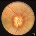
| 230 |
 |
Bilateral Severe Hemorrhagic Papilledema | Left eye. Bilateral Severe Hemorrhagic Papilledema in a woman with hyperthyroidism and dural sinus occlusion. | Image |
| 231 |
 |
Bilateral Severe Hemorrhagic Papilledema | Left eye. Bilateral hyperacute papilledema with rapid blindess associated with dural sinus occlusion. Both eyes were nearly blind. Boy. | Image |
| 232 |
 |
Buried and Visible Drusen | PP_19b: right eye : visible drusen in an eleven year old girl; PP_19a: left eye with buried drusen. Anatomy: Optic disc Pathology: Drusen of the optic disc Disease/Diagnosis: Drusen of the optic disc Clinical: Normally functioning eye with drusen. | Image |
| 233 |
 |
Buried and Visible Drusen | PP_19a Left eye with buried drusen. PP_19b: right eye : visible drusen. Eleven year old girl. Anatomy: Optic disc. Pathology: Drusen of the optic disc. Disease/Diagnosis: Drusen of the optic disc. Clinical notes: Normally functioning eye with drusen. | Image |
| 234 |
 |
Buried Drusen | 5 year old boy. Bilateral buried drusen. Notice the lumpy nasal disc elevation. This patient had a twin brother whose optic disc drusen were exposed. Anatomy: Optic disc. Pathology: Drusen of the optic disc. Disease/Diagnosis: Drusen of the optic disc. Clinical notes: Normally functioning eye with ... | Image |
| 235 |
 |
Buried Drusen | 5 year old boy. Bilateral buried drusen. Notice the lumpy nasal disc elevation. This patient had a twin brother whose optic disc drusen were exposed. Anatomy: Optic disc. Pathology: Drusen of the optic disc. Disease/Diagnosis: Drusen of the optic disc. Clinical notes: Normally functioning eye with ... | Image |
| 236 |
 |
Buried Drusen | 7 year old boy with pseudo papilledema from buried drusen. Note the lumpy contour of the disc margin. Also note the surrounding ring-like light reflex that is optically perfect and indicates absence of edema spreading onto the surrounding retina. Anatomy: Optic disc. Pathology: Drusen of the optic d... | Image |
| 237 |
 |
Buried Drusen | Young woman with pseudo papilledema from buried drusen with associated visual field defects. Barely visible in the upper arcuate nerve fibers is a slit like defect. Anatomy: Optic disc. Pathology: Drusen of the optic disc. Disease/Diagnosis: Drusen of the optic disc. Clinical notes: This patient had... | Image |
| 238 |
 |
Buried Drusen | Buried drusen with peculiar white dot, which appears to be choroidal in location. Note lumpy disc margin on right disc PP_15a is right eye. PP_15b is left eye. Beautiful example of pseudo papilledema in which drusen can not be seen. 8 year old girl. Anatomy: Optic disc. Pathology: Drusen of the op... | Image |
| 239 |
 |
Buried Drusen | Suspected buried drusen in a girl. Anatomy: Optic disc. Pathology: Drusen of the optic disc. Disease/Diagnosis: Drusen of the optic disc. Clinical notes: Normally functioning eye with suspected drusen. | Image |
| 240 |
 |
Buried Drusen | Left disc has a blurred lumpy margin. Retinal vessels are not obscured in the disc margin blur, therefore no edema is present. This is an example of a difficult blurred disc, the nature of which is clarified by the presence of a clear cut disk anomoly in the fellow eye. 8 year old girl. PP_15a has b... | Image |
| 241 |
 |
Buried Drusen | Buried drusen; PP_13a: Right eye. Note lumpy disc margin, especially temporally. Also note absence of optic cup. Excellent example of pseudo papilledema with buried drusen. Anatomy: Optic disc. Pathology: Drusen of the optic disc. Disease/Diagnosis: Drusen of the optic disc. Clinical notes: Patient ... | Image |
| 242 |
 |
Buried Drusen | Buried drusen. Left eye. Note lumpy disc margin, especially temporally. Also note absence of optic cup. Excellent example of pseudo papilledema with buried drusen. Pair with PP_13a. Anatomy: Optic disc. Pathology: Drusen of the optic disc. Disease/Diagnosis: Drusen of the optic disc. Clinical notes... | Image |
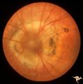
| 243 |
 |
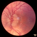
Buried Drusen with Choroidal Retinal Scar | Right eye: Buried drusen; probable complication of peripapillary hemorrhage at 7:00. Anatomy: Optic disc. Pathology: Drusen of the optic disc. Disease/Diagnosis: Drusen of the optic disc. Clinical notes: Enlarged blind spot. | Image |
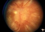
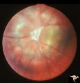
| 244 |
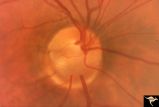 |
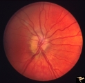
C01 Pits of the Optic Disc | Right eye. Very large inferior temporal optic pit. Congenital. Woman. Anatomy: Optic disc. | Image |
| 245 |
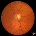 |
C03 Pits of the Optic Disc | Central optic pit. Left eye. Anatomy: Optic disc. | Image |
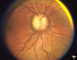
| 246 |
 |
C04 Pits of the Optic Disc | Right eye. Man. Large temporal pit. Macular detachment. Anatomy: Optic disc. | Image |
| 247 |
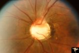 |
C08 Pits of the Optic Disc | Left eye. Large cavitary anomaly (pit). Man. 20/100 visual acuity. Superior nasal visual field defect. May not have a central retinal artery. Anatomy: Optic disc. Clinical: Man. 20/100 visual acuity. Superior nasal visual field defect. | Image |
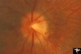
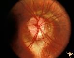
| 248 |
 |
C12 Morning Glory Disc | "Morning Glory" disc. Patient 11 year old. Anatomy: Optic disc. | Image |
| 249 |
 |
C16 Morning Glory Disc | "Morning Glory" disc. Note tapering edge pointing to basal encephalocele. Boy. Anatomy: Optic disc. | Image |
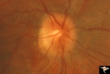
| 250 |
 |
C17 Morning Glory Disc | "Morning Glory" disc. CT normal. Anatomy: Optic disc. Clinical: CT normal. | Image |