A collection of videos relating to the diagnosis and treatment of eye movement disorders. This collection includes many demonstrations of examination techniques.
Dan Gold, D.O., Associate Professor of Neurology, Ophthalmology, Neurosurgery, Otolaryngology - Head & Neck Surgery, Emergency Medicine, and Medicine, The Johns Hopkins School of Medicine.
A collection of videos relating to the diagnosis and treatment of eye movement disorders.
NOVEL: https://novel.utah.edu/
TO
Filters: Collection: "ehsl_novel_gold"
| Title | Description | Type | ||
|---|---|---|---|---|
| 226 |
 |
Evaluation of Convergence | 𝗢𝗿𝗶𝗴𝗶𝗻𝗮𝗹 𝗗𝗲𝘀𝗰𝗿𝗶𝗽𝘁𝗶𝗼𝗻: The assessment of convergence includes measuring alignment at near versus distance (see video, https://collections.lib.utah.edu/details?id=187677), near point of convergence and convergence amplitude. Near point of conve... | Image/MovingImage |
| 227 |
 |
Optokinetic Nystagmus | 𝗢𝗿𝗶𝗴𝗶𝗻𝗮𝗹 𝗗𝗲𝘀𝗰𝗿𝗶𝗽𝘁𝗶𝗼𝗻: During the bedside evaluation of optokinetic nystagmus (OKN), the patient is instructed to look at each red (or white) square as it moves past. Because this is not a full-field visual stimuli, using an optokinetic flag m... | Image/MovingImage |
| 228 |
 |
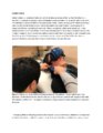
Pressure Testing for Superior Canal Dehiscence Syndrome | 𝗢𝗿𝗶𝗴𝗶𝗻𝗮𝗹 𝗗𝗲𝘀𝗰𝗿𝗶𝗽𝘁𝗶𝗼𝗻: Superior semicircular canal dehiscence syndrome (SCDS) is caused by a third mobile window in the inner ear. This allows for transmission of sound or pressure to the superior canal. Tragal compression and/or glottic and ... | Image/MovingImage |
| 229 |
 |
Provocative Maneuvers (Removal of Fixation, Vibration, Head-Shaking) to Accentuate Peripheral Vestibular Nystagmus) | 𝗢𝗿𝗶𝗴𝗶𝗻𝗮𝗹 𝗗𝗲𝘀𝗰𝗿𝗶𝗽𝘁𝗶𝗼𝗻: With an acute destructive process like vestibular neuritis that causes significant unilateral vestibular loss, spontaneous nystagmus is always present. However, over days to months, spontaneous nystagmus should resolve co... | Image/MovingImage |
| 230 |
 |
Evaluation of Auditory Function Using Rinne and Weber Tests | 𝗢𝗿𝗶𝗴𝗶𝗻𝗮𝗹 𝗗𝗲𝘀𝗰𝗿𝗶𝗽𝘁𝗶𝗼𝗻: The Rinne test is an assessment of auditory thresholds to air and bone conduction of sound. The Weber test is a comparison of bone conducted sound of either ear. Conductive hearing loss results in a loss of air conducte... | Image/MovingImage |
| 231 |
 |
Video Head Impulse Testing | 𝗢𝗿𝗶𝗴𝗶𝗻𝗮𝗹 𝗗𝗲𝘀𝗰𝗿𝗶𝗽𝘁𝗶𝗼𝗻: The video head impulse test (vHIT) is a clinical assessment technique used to assess the function of the semicircular canals-the angular acceleration detectors-which initiate the vestibulo-ocular reflex (VOR). The HIT and... | Text |
| 232 |
 |
ENG, VNG, & VOG | 𝗢𝗿𝗶𝗴𝗶𝗻𝗮𝗹 𝗗𝗲𝘀𝗰𝗿𝗶𝗽𝘁𝗶𝗼𝗻: Electronystagmography (ENG), and videonystagmography (VNG) or videooculography (VOG) are a collection of tests of eye movements that are performed either using surface electrodes around the eye (ENG) or with video goggles... | Text |
| 233 |
 |
Vestibular Evoked Myogenic Potentials (VEMPs) | 𝗢𝗿𝗶𝗴𝗶𝗻𝗮𝗹 𝗗𝗲𝘀𝗰𝗿𝗶𝗽𝘁𝗶𝗼𝗻: Vestibular-evoked myogenic potentials (VEMP) are electromyographic potential reflex tests that reflect the function of the saccule in cervical VEMP and the utricle in ocular VEMP.1 In the cervical VEMP an inhibitory refle... | Text |
| 234 |
 |
Caloric Testing | 𝗢𝗿𝗶𝗴𝗶𝗻𝗮𝗹 𝗗𝗲𝘀𝗰𝗿𝗶𝗽𝘁𝗶𝗼𝗻: Caloric testing is a peripheral vestibular test which takes advantage of the fact that the labyrinth is sensitive to temperature changes. Warm stimulation causes excitation of the semicircular canals while cold stimulatio... | Image/MovingImage |
| 235 |
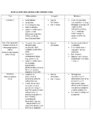 |
Summary of the Most Common Audio-Vestibular Testing | Chart describing common audio-vestibular testing. | Text |
| 236 |
 |
Audiometry | 𝗢𝗿𝗶𝗴𝗶𝗻𝗮𝗹 𝗗𝗲𝘀𝗰𝗿𝗶𝗽𝘁𝗶𝗼𝗻: Audiometry is the measurement of the sensitivity and range of an individual's hearing. As many etiologies of imbalance, nystagmus, vertigo and/or dizziness can have an otologic origin the audiogram is an important piece o... | Text |
| 237 |
 |
Rotary Chair Testing | 𝗢𝗿𝗶𝗴𝗶𝗻𝗮𝗹 𝗗𝗲𝘀𝗰𝗿𝗶𝗽𝘁𝗶𝗼𝗻: Rotary chair testing includes rotation around a vertical axis, and evaluates the horizontal semicircular canal vestibulo-ocular reflex (VOR). The patient sits in a mechanized chair with the head secured in a neutral posi... | Text |
| 238 |
 |
Sagging Eye Syndrome and Cerebellar Disease in Divergence Insufficiency | 𝗢𝗿𝗶𝗴𝗶𝗻𝗮𝗹 𝗗𝗲𝘀𝗰𝗿𝗶𝗽𝘁𝗶𝗼𝗻: This is a 70-year-old woman who presented with diplopia at distance. Her exam demonstrated orthophoria at near with a fairly comitant 8-10 PD esotropia at distance without abduction paresis, consistent with divergence ins... | Image/MovingImage |
| 239 |
 |
HINTS Exam and Saccadic Dysmetria in Lateral Medullary Stroke | 𝗢𝗿𝗶𝗴𝗶𝗻𝗮𝗹 𝗗𝗲𝘀𝗰𝗿𝗶𝗽𝘁𝗶𝗼𝗻: This is a 50-year-old who experienced the abrupt onset of prolonged vertigo following chiropractic therapy 2 months prior. Initial work-up included an MRI and MR angiogram - MR-diffusion weighted imaging showed an acute l... | Image/MovingImage |
| 240 |
 |
Medial Longitudinal Fasciculus Syndrome with Prominent Spontaneous Nystagmus | 𝗢𝗿𝗶𝗴𝗶𝗻𝗮𝗹 𝗗𝗲𝘀𝗰𝗿𝗶𝗽𝘁𝗶𝗼𝗻: This is a 60-year-old man who experienced the abrupt onset of diplopia and imbalance. He had typical features of a left medial longitudinal fasciculus (MLF) syndrome including left internuclear ophthalmoplegia (INO) and l... | Image/MovingImage |
| 241 |
 |
Isolated Central 4th Nerve Palsy | 𝗢𝗿𝗶𝗴𝗶𝗻𝗮𝗹 𝗗𝗲𝘀𝗰𝗿𝗶𝗽𝘁𝗶𝗼𝗻: This is a 40-year-old man with a right hypertropia that worsened in left and down gaze in addition to right head tilt, and improved in left head tilt. There was subjective excyclotorsion OD with double Maddox rod testing.... | Image/MovingImage |
| 242 |
 |
Torsional Nystagmus Due to Medullary Pilocytic Astrocytoma | 𝗢𝗿𝗶𝗴𝗶𝗻𝗮𝗹 𝗗𝗲𝘀𝗰𝗿𝗶𝗽𝘁𝗶𝗼𝗻: This is a 30-year-old woman who experienced headaches which led to an MRI and the diagnosis of a right medullary pilocytic astrocytoma, confirmed pathologically. Examination was performed a year after the initial diagnosi... | Image/MovingImage |
| 243 |
 |
Head-Shaking Nystagmus - A 'Central' Pattern | 𝗢𝗿𝗶𝗴𝗶𝗻𝗮𝗹 𝗗𝗲𝘀𝗰𝗿𝗶𝗽𝘁𝗶𝗼𝗻: Evaluating for nystagmus provoked by head-shaking, so-called head-shaking nystagmus (HSN), should be performed in all patients with complaints of dizziness or vertigo, regardless of the chronicity. The maneuver is perform... | Image/MovingImage |
| 244 |
 |
Downbeat Nystagmus | 𝗢𝗿𝗶𝗴𝗶𝗻𝗮𝗹 𝗗𝗲𝘀𝗰𝗿𝗶𝗽𝘁𝗶𝗼𝗻: This is a 40-year-old man with 2 years of progressive ataxia and oscillopsia. On examination, he had downbeat nystagmus (DBN), an ocular motor finding that is usually (but not always) associated with flocculus/parafloccul... | Image/MovingImage |
| 245 |
 |
Peripheral (Vestibular) and Central (Gaze-Evoked) Patterns of Nystagmus in a Single Patient | A 55-year-old man experienced episodic vertigo and was diagnosed with Meniere's disease affecting the left ear (based on audiograms and his clinical course) about 1 year prior to presentation. About 6 months prior to presentation, intratympanic (IT) gentamicin was injected into the left ear, at whic... | Image/MovingImage |
| 246 |
 |
Central HINTS (With an Abnormal Head Impulse Sign) in the Acute Vestibular Syndrome Due to Lateral Pontine/Middle Cerebellar Peduncle Demyelination | 𝗢𝗿𝗶𝗴𝗶𝗻𝗮𝗹 𝗗𝗲𝘀𝗰𝗿𝗶𝗽𝘁𝗶𝗼𝗻: This is a 30-year-old man presenting with vertigo, diplopia and mild left facial weakness (not seen in the video). On exam, there was right-beating nystagmus (RBN) in primary gaze that increased in right gaze (in accordan... | Image/MovingImage |
| 247 |
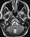 |
Oculopalatal Tremor with Prominent Nystagmus, Bilateral Horizontal Gaze Palsy, and Bilateral Facial Palsies (Figure 1) | Figure 1, MRI T2 sequence demonstrating hyperintensities involving bilateral inferior olives of the medulla. This is a 50-year-old woman who experienced the acute onset of right sixth and seventh nerve palsies and left hemiparesis. Two cavernomas within the right pons (one in the region of the facia... | Image |
| 248 |
 |
Skew Deviation and Spontaneous Nystagmus Due to Posterior Fossa Lesions | This is a 50-year-old woman who reported the abrupt onset of imbalance, right upper extremity incoordination and binocular vertical diplopia several months prior to her presentation to our clinic. On examination, she had a left hypertropia that was fairly comitant (measuring 5 prism diopters) assoc... | Image/MovingImage |
| 249 |
 |
Oculopalatal Tremor with Prominent Nystagmus, Bilateral Horizontal Gaze Palsy, and Bilateral Facial Palsies | 𝗢𝗿𝗶𝗴𝗶𝗻𝗮𝗹 𝗗𝗲𝘀𝗰𝗿𝗶𝗽𝘁𝗶𝗼𝗻: This is a 50-year-old woman who experienced the acute onset of right sixth and seventh nerve palsies and left hemiparesis. Two cavernomas within the right pons (one in the region of the facial colliculus) were demonstrat... | Image/MovingImage |
| 250 |
 |
Slow Volitional Saccades and Poor Fast Phases to an Optokinetic Stimulus, with Preserved Head Impulse Testing | This is a 67-year-old woman presenting with imbalance and binocular horizontal diplopia at near. On examination there were frequent square wave jerks, limited supraduction OU and convergence insufficiency, which explained her diplopia. Pursuit and suppression of the vestibulo-ocular reflex were sa... | Image/MovingImage |