Best known for his world-renowned neuro-ophthalmology unit based at the University of California, San Francisco, William Hoyt, MD collected here more than 850 of his best images covering a wide range of disorders.
William F. Hoyt, MD, Professor Emeritus of Ophthalmology, Neurology and Neurosurgery, Department of Ophthalmology, University of California, San Francisco.
NOVEL: https://novel.utah.edu/
TO
| Title | Description | Type | ||
|---|---|---|---|---|
| 226 |
 |
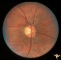
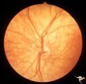
H09 Panhypoplasia | Moderate hypoplasia. Man. Anatomy: Optic disc. Pathology: Hypoplasia of the optic nerve. Disease/ Diagnosis: Hypoplasia. | Image |
| 227 |
 |
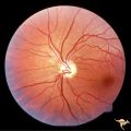
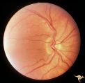
H10 Panhypoplasia | Cruzon's Disease. 26 year old man. Right eye. Mild hypoplasia. Son of patient in H_11 and H_12. Same patient in H_31. Father of patient in H_32. Anatomy: Optic disc. Pathology: Hypoplasia of the optic nerve. Disease/ Diagnosis: Hypoplasia. | Image |
| 228 |
 |
H101 Occipital Hemianoptic Hypoplasia | Right eye. Same patient as H_102. Anatomy: Optic disc. Pathology: Occipital hemianoptic hypoplasia. Disease/ Diagnosis: Congenital defect of the occipital lobe. | Image |
| 229 |
 |
H102 Occipital Hemianoptic Hypoplasia | Left eye. Trans-synaptic band atrophy. Left homonymous hemianopia from right occipital porencephaly. Loss of nasal nerve fibers. Same patient as H_101. Anatomy: Optic disc. Pathology: Occipital hemianoptic hypoplasia. Disease/ Diagnosis: Congenital defect of the occipital lobe. | Image |
| 230 |
 |
H103 Occipital Hemianoptic Hypoplasia | Right eye. Congenital right homonymous hemianopia. Absent nerve fiber layer in right eye. Same patient as H_104. Anatomy: Optic disc. Pathology: Occipital hemianoptic hypoplasia. Disease/ Diagnosis: Congenital defect of the occipital lobe. | Image |
| 231 |
 |
H104 Occipital Hemianoptic Hypoplasia | Left eye. Contrast with nasal nerve fiber in right eye, H_103. Anatomy: Optic disc. Pathology: Occipital hemianoptic hypoplasia. Disease/ Diagnosis: Congenital defect of the occipital lobe. | Image |
| 232 |
 |
H105 Occipital Hemianoptic Hypoplasia | Left congenital homonymous hemianopia. Right occipital AVM. Nasal nerve fiber layer loss in left eye. Compare with right eye. Same patient as H_106. Anatomy: Optic disc. Pathology: Occipital hemianoptic hypoplasia. DIsease/ Diagnosis: Congenital defect of the occipital lobe | Image |
| 233 |
 |
H106 Occipital Hemianoptic Hypoplasia | Same patient as H_105. Anatomy: Optic disc. Pathology: Occipital hemianoptic hypoplasia. Disease/ Diagnosis: Congenital defect of the occipital lobe. | Image |
| 234 |
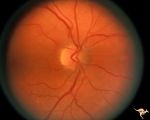 |
H11 Panhypoplasia | Cruzon's Disease. 47 year old woman. Right eye. Mild hypoplasia. Mother of patient in H_10 and H_31. Same patient as H_12. Grandmother of patient in H_32. Anatomy: Optic disc. Pathology: Hypoplasia of the optic nerve. Disease/ Diagnosis: Hypoplasia. | Image |
| 235 |
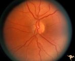 |
H12 Panhypoplasia | Cruzon's Disease. 47 year old woman. Left eye. Mild hypoplasia. Mother of patient in H_10 and H_31. Same patient as H_11. Grandmother of patient in H_32. Anatomy: Optic disc. Pathology: Hypoplasia of the optic nerve. Pathology: Hypoplasia of the optic nerve. Disease/ Diagnosis: Hypoplasia. | Image |
| 236 |
 |
H13 Panhypoplasia | Right eye. Blind baby. Severe hypoplasia with blond fundus. Same patient as H_14. Anatomy: Optic disc. Pathology: Hypoplasia of the optic nerve. Disease/ Diagnosis: Hypoplasia. Imaging: Hypoplasia of the optic nerve. | Image |
| 237 |
 |
H14 Panhypoplasia | Left eye. Blind baby. Severe hypoplasia with blond fundus. Same patient as H_13. Anatomy: Optic disc. Pathology: Hypoplasia of the optic nerve. Disease/ Diagnosis: Hypoplasia. | Image |
| 238 |
 |
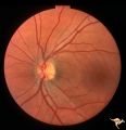
H15 Panhypoplasia | Moderate hypoplasia. Right eye. 14 year old boy. Good example of double ring sign. Same patient as H_16. Anatomy: Optic disc. Pathology: Hypoplasia of the optic nerve. Disease/ Diagnosis: Hypoplasia. | Image |
| 239 |
 |
H16 Panhypoplasia | Moderate hypoplasia. Left eye. 14 year old boy. Good example of double ring sign. Same patient as H_15. Anatomy: Optic disc. Pathology: Hypoplasia of the optic nerve. Disease/ Diagnosis: Hypoplasia. | Image |
| 240 |
 |
H17 Panhypoplasia | Bilateral mild hypoplasia without field defect. Right eye. 30 year old woman. Same patient as H_18. Anatomy: Optic disc. Pathology: Hypoplasia of the optic nerve. Disease/ Diagnosis: Hypoplasia. | Image |
| 241 |
 |
H18 Panhypoplasia | Bilateral mild hypoplasia without field defect. Left eye. 30 year old woman. Same patient as H_17. Anatomy: Optic disc. Pathology: Hypoplasia of the optic nerve. Disease/ Diagnosis: Hypoplasia. | Image |
| 242 |
 |
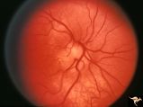
H21 Panhypoplasia | Right eye. Hypoplasia with glial tissue haze. Same patient as H_22. Anatomy: Optic disc. Pathology: Hypoplasia of the optic nerve. Disease/ Diagnosis: Hypoplasia. | Image |
| 243 |
 |
H22 Panhypoplasia | Left eye. Normal disc. Same patient as H_21. Anatomy: Optic disc. Pathology: Hypoplasia of the optic nerve. Disease/ Diagnosis: Hypoplasia. | Image |
| 244 |
 |
H27 Dysplasia with Hypoplasia (Elevated Dysplasia with Anomalous Vessels) | Right eye. Elevated hypoplastic dysplasia with anomalous vessels. Same patient as H_28. Anatomy: Optic disc. Pathology: Dysplasia of the optic disc. Disease/ Diagnosis: Elevated dysplasia with hypoplasia. | Image |
| 245 |
 |
H28 Dysplasia with Hypoplasia (Elevated Dysplasia with Anomalous Vessels) | Left eye. Elevated hypoplastic dysplasia with tortuous anomalous vessels. Same patient as H_27. Anatomy: Optic disc. Pathology: Dyplasia of the optic disc. Disease/ Diagnosis: Elevated dysplasia with hypoplasia. | Image |
| 246 |
 |
H29 Dysplasia with Hypoplasia (Elevated Dysplasia with Anomalous Vessels) | Right eye. Dysplasia with anomalous cilioretinal arterioles. Central retinal artery may be absent. Same patient as H_30. Anatomy: Optic disc. Pathology: Dysplasia of the optic disc. Disease/ Diagnosis: Elevated dysplasia with hypoplasia. | Image |
| 247 |
 |
H30 Dysplasia with Hypoplasia (Elevated Dysplasia with Anomalous Vessels) | Left eye. Elevated dysplasia with anomalous cilioretinal vessels. Central retinal artery may be absent. Same patient as H_29. Anatomy: Optic disc. Pathology: Dysplasia of the optic disc. Disease/ Diagnosis: Elevated dysplasia with hypoplasia. | Image |
| 248 |
 |
H53 Superior Segmental Optic Hypoplasia (SSOH) Topless Disc Syndrome | Superior segmental optic hypoplasia. High exit point of central retinal vessels. Anatomy: Optic disc. Pathology: Superior segmental optic hypoplasia (SSOH). Disease/ Diagnosis: Superior segmental optic hypoplasia (SSOH). | Image |
| 249 |
 |
H54 Superior Segmental Optic Hypoplasia (SSOH) Topless Disc Syndrome | Note superior segmental palor. Anatomy: Optic disc. Pathology: Superior segmental optic hypoplasia (SSOH). Disease/ Diagnosis: Congenital anomaly. | Image |
| 250 |
 |
H55 Superior Segmental Optic Hypoplasia (SSOH) Topless Disc Syndrome | Note superior choroidal crescent. Anatomy: Optic disc. Pathology: Superior segmental optic hypoplasia (SSOH). Disease/ Diagnosis: Congenital anomaly. | Image |