Best known for his world-renowned neuro-ophthalmology unit based at the University of California, San Francisco, William Hoyt, MD collected here more than 850 of his best images covering a wide range of disorders.
William F. Hoyt, MD, Professor Emeritus of Ophthalmology, Neurology and Neurosurgery, Department of Ophthalmology, University of California, San Francisco.
NOVEL: https://novel.utah.edu/
TO
| Title | Description | Type | ||
|---|---|---|---|---|
| 226 |
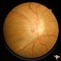 |
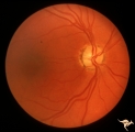
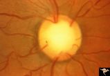
Post Papilledema Retinal Choroidal Bypass (Optociliary) | Right eye. Post papilledema retinal choroidal bypass (optociliary). Arterial venous malformation draining into saggital sinus causing papilledema and retinal choroidal collaterals. Anatomy: Optic disc. Pathology: Post papilledema. Disease/Diagnosis: Post papilledema with retinal choroidal bypass ves... | Image |
| 227 |
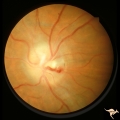 |
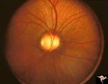
Post Papilledema Retinal Choroidal Bypass (Optociliary) | Left eye. Post papilledema retinal choroidal bypass (optociliary). Arterial venous malformation draining into saggital sinus causing papilledema and retinal choroidal collaterals. Anatomy: Optic disc. Pathology: Post papilledema. Disease/Diagnosis: Post papilledema with retinal choroidal bypass vess... | Image |
| 228 |
 |
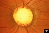
IC102c Central Retinal Artery Occlusion with Cilioretinal Collaterals | Right eye, 1982, Central retinal artery occlusion with cilioretinal collateral occlusions due to calcific embolic occlusion behind the lamina cribrosa due to calcific valvular heart disease. Collaterals have been called "Nettleship Collaterals", recognizing the British physician who first described ... | Image |
| 229 |
 |
IC102a Central Retinal Artery Occlusion with Cilioretinal Collaterals | Left eye, 1988, Central retinal artery with cilioretinal collaterals due to calcific embolic behind the lamina cribrosa due to calcific valvular heart disease. Collaterals have been called "Nettleship Collaterals", recognizing the British physician who first described them in 1892. Anatomy: Optic di... | Image |
| 230 |
 |
IC102b Central Retinal Artery Occlusion with Cilioretinal Collaterals | Right eye, 1991, Central retinal artery occlusion with cilioretinal collateral occlusions due to calcific embolic occlusion behind the lamina cribrosa due to calcific valvular heart disease. Collaterals have been called "Nettleship Collaterals", recognizing the British physician who first described ... | Image |
| 231 |
 |
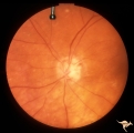
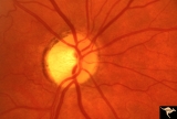
E02 Disc Swelling with Central Vein Occlusion | 37 year old black male with sickle cell C causing unilateral central retinal vien occlusion. Anatomy: Optic disc; Retina. Pathology: Occlusion of the central retinal vein. Disease/ Diagnosis: Disc swelling due to central retial vein occlusion. Clinical: Visual blurring. | Image |
| 232 |
 |
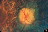
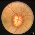
IF107 Glaucoma Cupped Disc | Glaucoma cupped disc with inferior temporal retinal nerve fiber layer defect. Vertically ovoid cup. 1974. Anatomy: Optic disc. Pathology: Glaucoma. Disease/ Diagnosis: Glaucoma. Clinical: Superior arcuate visual field defects. | Image |
| 233 |
 |
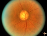
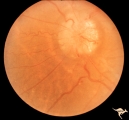
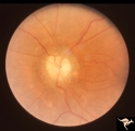
Visible Drusen with Retinitis Pigmentosa | Right eye. Optic disc drusen with retinitis pigmentosa. Note the marked narrowing of the retinal arterioles and the spectacular change in the peripapillary choroid. Anatomy: Optic disc. Pathology: Drusen of the optic disc. Disease/Diagnosis: Drusen of the optic disc. Clinical: Patient was nearly bli... | Image |
| 234 |
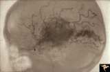 |
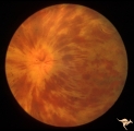
Vascular Disc Anomalies - Retinal Arteriovenous Malformations | Retinal arteriovenous malformations. Wyburn-Mason Syndrome. Angiogram showing extension of vascular malformation up the right optic nerve (arrow) through the thalamus and into the right visual cortex. References #3 and #73. Anatomy: Brain. Pathology: Arteriovenous malformation. Disease/Diagnosis: Wy... | Image |
| 235 |
 |
Segmental Atrophy - Hemianopic (Band) Atrophy | Segmental Atrophy - Band atrophy in an eye with temporal hemianopia. Wyburn-Mason Syndrome extending to the chiasm. Left eye 1975. Anatomy: Optic disc. Pathology: Right sided chiasmal AVM. Disease/Diagnosis: Band atrophy due to chiasmal AVM and Wyburn-Mason Syndrome. Clinical: Blind right eye, temp... | Image |
| 236 |
 |
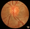
IE02b Acute Leber Optic Neuropathy | Acute Leber Optic Neuropathy. Formation of hemorrhage one year after IE_02a. At this time, he was beginning to lose central vision. .Note thinning of the nerve fiber layer temporally, 3:00 - 4:00 Pair with IE_02a. March 26, 1980. Anatomy: Optic disc. Pathology: Optic neuropathy. Disease/ Diagnosis: ... | Image |
| 237 |
 |
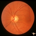
IE01 Acute Leber Optic Neuropathy | Pseudo edema with peripapillary microangiopathy in a brother of boy with Leber Optic Neuropathy. Not pale nerve yet. Right eye. Same patient as IE_02a and IE_02b. August 7, 1979. Anatomy: Optic disc. Pathology: Optic neuropathy. Disease/ Diagnosis: Optic neuropathy. Clinical: Asymptomatic | Image |
| 238 |
 |
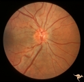
IE02a Acute Leber Optic Neuropathy | Acute Leber Optic Neuropathy, Left eye. Same patient as IE_01 and IE_02b. August 7, 1979. Anatomy: Optic disc. Pathology: Optic neuropathy. Disease/ Diagnosis: Leber's optic neuropathy. Clinical: Asymptomatic. | Image |
| 239 |
 |
IF201a Temporal Cupping with Dominant Hereditary Optic Atrophy | 1969. Dominant hereditary optic atrophy (Kjer) Pair with IF2_1b. Right eye. Boy with reduced central acuity since childhood. Discs are pale temporally and the temporal nerve fiber layer is thin. Anatomy: Optic disc. Pathology: Dominant hereditary optic atrophy. Disease/ Diagnosis: Dominant hereditar... | Image |
| 240 |
 |
IF205a Temporal Cupping with Dominant Hereditary Optic Atrophy | 1970. Right eye. Pair with IF2_5b. 55 year old woman with deficient vision all her life. Typical pattern of dominant hereditary atrophy. Temporal pallor and shallow cupping. Anatomy: Optic disc. Pathology: Dominant hereditary optic atrophy. Disease/ Diagnosis: Dominant hereditary optic atrophy. Cli... | Image |
| 241 |
 |
IF205b Temporal Cupping with Dominant Hereditary Optic Atrophy | 1970. Left eye. Pair with IF2_5a. 55 year old woman with deficient vision all her life. Typical pattern of dominant hereditary atrophy. Temporal pallor and shallow cupping. Anatomy: Optic disc. Pathology: Dominant hereditary optic atrophy. Disease/ Diagnosis: Dominant hereditary optic atrophy. Clin... | Image |
| 242 |
 |
IF201b Temporal Cupping with Dominant Hereditary Optic Atrophy | 1969. Dominant hereditary optic atrophy (Kjer) Pair with IF2_1a. Left eye. Boy with reduced central acuity since childhood. and the temporal nerve fiber layer is thin. Anatomy: Optic disc. Pathology: Dominant hereditary optic atrophy. Disease/ Diagnosis: Dominant hereditary optic atrophy. Clinical: ... | Image |
| 243 |
 |
C23 Empty Disc | Father of patient in C_21 and C_22. Father has central retinal artery, multiple cilioretinal arteries and had previously unsuspected renal failure. Papillorenal Syndrome (PRS). Reference: Parsa,CF et al. Ophthalmology. 2001. 108(4): 738-49Barroso LH, Hoyt WF, Narahara M. Can the arterial supply of ... | Image |
| 244 |
 |
C21 Empty Disc | Right eye. All cilioretinal fundus. No central retinal artery. Handmann anomaly. Frequently associated with renal dysplasia. Pair with C_22 an C_23. Reference: Barroso LH, Hoyt WF, Narahara M. Can the arterial supply of the retina in man be exclusively cilioretinal? J Neuroophthalmol. 1994 Jun;14(2... | Image |
| 245 |
 |
C22 Empty Disc | Left eye. All cilioretinal fundus. No central retinal artery. Handmann anomaly. Frequently associated with renal dysplasia. Pair with C_21 an C_23. Reference: Barroso LH, Hoyt WF, Narahara M. Can the arterial supply of the retina in man be exclusively cilioretinal? J Neuroophthalmol. 1994 Jun;14(2)... | Image |
| 246 |
 |
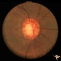
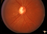
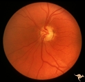
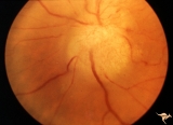
Buried Drusen | 7 year old boy with pseudo papilledema from buried drusen. Note the lumpy contour of the disc margin. Also note the surrounding ring-like light reflex that is optically perfect and indicates absence of edema spreading onto the surrounding retina. Anatomy: Optic disc. Pathology: Drusen of the optic d... | Image |
| 247 |
 |
Chronic Papilledema with Pseudo Drusen | Right eye. Meningioma. Pseudo drusen from chronic papilledema. Woman. Anatomy: Optic disc Pathology: Papilledema Disease/Diagnosis: Chronic papilledema with pseudo drusen | Image |
| 248 |
 |
Chronic Papilledema with Pseudo Drusen | Left eye. Meningioma. Pseudo drusen from chronic papilledema. The patient's meningioma had blinded her left eye and caused chronic elevated intracranial pressure. Woman. Anatomy: Optic disc Pathology: Papilledema Disease/Diagnosis: Chronic papilledema with pseudo drusen | Image |
| 249 |
 |
F107 Metastatic Breast Cancer to the Disc | Metastatic breast cancer to the disc. Notice mass on inferior portion of disc. Also notice tangled capillary dilation within the mass indicating infiltration. This disc tumor was radiated. It disappeared leaving a pale flat atrophic nerve. The patient died. Histologic study of the eye revealed metas... | Image |
| 250 |
 |
Buried Drusen with Choroidal Retinal Scar | Right eye: Buried drusen; probable complication of peripapillary hemorrhage at 7:00. Anatomy: Optic disc. Pathology: Drusen of the optic disc. Disease/Diagnosis: Drusen of the optic disc. Clinical notes: Enlarged blind spot. | Image |