Collection of materials relating to neuro-ophthalmology as part of the Neuro-Ophthalmology Virtual Education Library.
NOVEL: https://novel.utah.edu/
TO
- NOVEL226
| Title | Creator | Description | Subject | ||
|---|---|---|---|---|---|
| 1 |
 |
Clinical Characteristics of Ocular Lateropulsion | Jorge C Kattah, MD | A discussion of the normal mechanism that maintain the eyes in normal horizontal position. | Ocular Lateropulsion; Unilateral Gaze Palsy; Radiographic h-CGD |
| 2 |
 |
Cogan's Lid Twitch Sign | Raed Behbehani, MD | Cogan's lid twitch sign is a twitch sign of he upper lid upon looking straight from a sustained downgaze position. It is associated with Ocular Myasthenia Gavis. | Myasthenia; Ptosis; Lid Twitch |
| 3 |
 |
Common Patterns of Visual Field Defects | Sean Gratton, MD; Sarah Lam, 6th year BA/MD | Lecture covering common visual field defects, including those of the retina, optic nerve, chiasm, and retrochiasmal. | Visual Field Defects |
| 4 |
 |
Complications of Strabismus Surgery and Botox | W. Walker Motley, MD | A narrated video slideshow outlining complications associated with strabismus surgery. | Strabismus; Surgery; Surgical Complications; Botox |
| 5 |
 |
Cranial Nerves: Neuroanatomy Video Lab - Brain Dissections | Suzanne S. Stensaas, PhD | The approach is to learn to associate the cranial nerves with their brainstem level and blood supply. Emphasis is given to the midbrain (3, 4), pons (5, 6, 7, 8), medulla (9, 10, 11, 12) and their most important functions. | Cranial Nerves; Brain; Dissection |
| 6 |
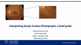 |
Interpreting Ocular Fundus Photographs: A Brief Guide | Gabriele Berman; Sachin Kedar; Nancy J. Newman; Valerie Biousse | This is a brief guide to the interpretation of the ocular fundus photograph. In this presentation we will describe the structures that comprise the normal ocular fundus followed the abnormalities that can be detected on fundus photographs. By the end of the presentation, learners should be able to d... | Fundus Photograph; Glaucoma; Papilledema; Retinal Detachment |
| 7 |
 |
A Five-Minute, Intravenous Edrophonium -Window to Observe the CNS Response to Myasthenic Ophthalmoplegia | Jorge C Kattah, MD | A video showing how ocular motor adaptation may be observed. | Ocular Motor Adaptation |
| 8 |
 |
Nystagmus Elicitation Techniques | Jorge C. Kattah, MD | An examination of the patient days or weeks after the acute event requires fixation block, and a variety of techniques, known as nystagmus elicitation maneuvers to detect the recent vestibular imbalance. | Nystagmus |
| 9 |
 |
Natural Language Processing in Neuro-Ophthalmology | Areeba Abid, BS; Sachin Kedar, MD | This video provides an overview of natural language processing (NLP) techniques, applications, and limitations in neuro-ophthalmology. NLP, a branch of artificial intelligence, enables computers to understand and analyze human language. This video discusses three types of NLP techniques: sentiment a... | Artificial Intelligence; Machine Learning; Natural Language Processing; Word Prediction; Word Embeddings; Sentiment Analysis |
| 10 |
 |
Upbeat Nystagmus | Raed Behbehani, MD, | A patient with a brain stem syndrome due to demyelination and upbeat nystagmus. | Upbeat Nystagmus |
| 11 |
 |
One and a Half Syndrome Following Resection of a Posterior Fossa Epidermoid Cyst | Christine Xu; Claire Basco, MS, NP-C; Kiarash Shahlaie; Yin Allison Liu | This is a case of One and a Half Syndrome following resection of a posterior fossa epidermoid cyst. A 31-year-old male initially presented with left facial droop and bilateral ptosis, and a "down and out" gaze of the left eye. He underwent imaging and was diagnosed with an epidermoid cyst located in... | Abducens Nucleus; Epidermoid Cyst; Extraocular Movements; Gaze Palsy; Internuclear Ophthalmoplegia; Medial Longitudinal Fasciculus; Neurosurgery; One and a Half Syndrome |
| 12 |
 |
Hypothalamus: Neuroanatomy Video Lab - Brain Dissections | Suzanne S. Stensaas, PhD | Gross specimens are used to demonstrate the area of the hypothalamus and its relationship to surrounding structures. Both endocrine and autonomic functions are explored using diagrams. Mention is made of the direct hypothalamic response to circulating hormones and other substances such as sodium. Th... | Hypothalamus; Brain; Dissection |
| 13 |
 |
Three Critical Vertical Pathways: Neuroanatomy Video Lab - Brain Dissections | Suzanne S. Stensaas, PhD | There is one motor and two sensory pathways that must be mastered. Pain and temperature from the body travel together and vibration and proprioception travel in another pathway each reaching perception in the cortex. Voluntary motor control starts in the cerebral cortex and connects with a motor neu... | Spinothalamic Tract; Dorsal Column-Medical Lemniscus Pathway; Posterior Column; Vertical Pathway; Brain; Dissection |
| 14 |
 |
The Spinal Cord & Monosynaptic Reflex: Neuroanatomy Video Lab - Brain Dissections | Suzanne S. Stensaas, PhD | The spinal cord's relationship to the foramina, discs and spinal nerves is demonstrated on a model. The dura, ganglia and rootlets are shown as well as the gray and white matter in gross sections at different levels. A model of the cord is used to demonstrate and describe the anatomy of a monosynapt... | Spinal Cord; Monosynaptic Reflex; Brain; Dissection |
| 15 |
 |
Limbic System: Neuroanatomy Video Lab - Brain Dissections | Suzanne S. Stensaas, PhD | The decision was made to present a simplified description of a much more complex system using animations to construct a 3D image. Papez circuit is shown on gross specimens with mention of its involvement in memory. The role of the amygdala in fear and the olfactory cortex in temporal lobe epilepsy a... | Limbic System; Hippocampus; Brain; Dissection |
| 16 |
 |
The Most Important Pathway: Motor Control: Neuroanatomy Video Lab - Brain Dissections | Suzanne S. Stensaas, PhD | The origin of the corticospinal tract in the cerebral cortex is traced through gross sections of the hemisphere and brain stem to the spinal cord. Using an animation, the terms upper and lower motor neuron are defined and clinical signs and symptom listed. | Corticospinal Tract; Cerebral Cortex; Motor Neuron; Brain; Dissection |
| 17 |
 |
The Unfixed Spinal Cord: Neuroanatomy Video Lab - Brain Dissections | Suzanne S. Stensaas, PhD | The spinal cord's relationship to the foramina, discs and spinal nerves is demonstrated on a model. The dura, ganglia and rootlets are shown as well as the gray and white matter in gross sections at different levels. A model of the cord is used to demonstrate and describe the anatomy of a monosynapt... | Unfixed Spinal Cord; Spinal Cord; Brain; Dissection |
| 18 |
 |
Olfactory System: Neuroanatomy Video Lab - Brain Dissections | Suzanne S. Stensaas, PhD | Beginning with the location of the sensory cells within the skull the axons are traced into the cranial cavity. Demonstration of the olfactory bulb, olfactory tract and it termination in the forebrain and temporal lobe are indicated. Trauma and meningiomas can produce loss of small (anosmia). Degene... | Olfactory System; Olfactory Bulb; Anosmia; Brain; Dissection |
| 19 |
 |
The Meninges: Neuroanatomy Video Lab - Brain Dissections | Suzanne S. Stensaas, PhD | The epidural, subdural and subarachnoid spaces are demonstrated and discussed with respect to trauma and disease. The relationship of the brainstem and cerebellum to the tentorium demonstrates the vulnerability of the brain stem to increased supratentorial pressure and herniation. Arachnoid granulat... | Meninges; Brain; Dissection |
| 20 |
 |
Orientation: The Planes of the Brain: Neuroanatomy Video Lab - Brain Dissections | Suzanne S. Stensaas, PhD | Terms such as anterior, posterior, inferior and superior are introduced with respect to the hemispheres as well as the brain stem. Terms such as rostral and caudal or dorsal and ventral can mean different things in different areas. Sections in three planes (frontal, axial, and sagittal) are demonstr... | Frontal; Axial; Sagittal; Brain; Dissection |
| 21 |
 |
The Visual Pathway: Neuroanatomy Video Lab - Brain Dissections | Suzanne S. Stensaas, PhD | A brief review of the anatomy of the eye and the photic stimulation of the receptors is followed by a gross exploration of the visual pathway from the optic nerve, chiasm, and tract to the thalamus stressing how the left part of the visual world reaches the right hemisphere. Visual fields are relate... | Visual Pathway; Brain; Dissections |
| 22 |
 |
The Normal Unfixed Brain: Neuroanatomy Video Lab - Brain Dissections | Suzanne S. Stensaas, PhD | The consistency and vulnerability of the brain is demonstrated along with the clear and glistening pia and arachnoid and the tough dura. The cushioning function of the CSF is stressed and the features are pointed out on the ventral surface. The uncus and temporal lobes are normal with arteries free ... | Brain; Dissections |
| 23 |
 |
Parinaud Syndrome | Raed Behbehani, MD | Parinaud syndrome, as called dorsal midbrain syndrome, is due to dorsal midbrain lesions from compression (e.g., a tumor), demyelination, or ischemia. The syndrome is characterized by limitation of upward gaze, convergence retraction nystagmus, light near dissociation, and lid retraction (Collier's ... | Dorsal Mibrain Syndrome; Parinaud's Syndrome |
| 24 |
 |
Square Wave Jerks with Contrapulsion | Raed Behbehani, MD | A patient with history of brain stem stroke 2 months ago (right hemifacial anesthesia , left sided weakness and bulbar symptoms dysphagia) comes complaining of oscillipsia , binocular vertical diplopia). On exam he had a vertical tropia of 3-4 PD (Skew deviation), dissociated nystagmus , and saccadi... | Square Wave Jerks; Contrapulsion |
| 25 |
 |
Oculopalatal Tremor | Raed Behbehani, MD | This is a usually vertical, pendular nystagmus associated with synchronous rhythmic movement of the palate, developing months after a severe brain stem stroke. The stroke involves the dentato-rubro-olivary tract (Mollaret's triangle). MRI can show hypertrophy of the inferior olivary nucleus in the m... | Oculopalatal Tremor |