AAO-NANOS Neuro-Ophthalmology Clinical Collection: Derived from the AAO-NANOS Clinical Neuro-Ophthalmology collection produced on CD. The images are of selected cases from the NANOS teaching slide exchange, and the CD was produced under the direction of Larry Frohman, MD and Andrew Lee, MD.
The American Academy of Ophthalmology (AAO); The North American Neuro-Ophthalmology Association (NANOS).
NOVEL: https://novel.utah.edu/
TO
1 - 25 of 2
| Title | Description | Subject | ||
|---|---|---|---|---|
| 1 |
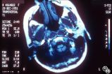 |
Neuro-Ophthalmic Imaging-MRI | Axial view of Arnold-Chiari malformation on a patient with downbeat nystagmus. Note the presence of the cerebellar tonsils posterior to the caudal medulla. In addition to downbeat nystagmus, Arnold-Chiari malformations can sometimes lead to increased intracranial pressure and papilledema. | Arnold-Chiari Malformation; Chiari Malformation; Inferior Tonsillar Herniation; Downbeat Nystagmus |
| 2 |
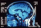 |
Neuro-Ophthalmic Imaging-MRI | Sagittal view of Arnold-Chiari malformation on a patient with downbeat nystagmus. The compression of the cervicomedullary junction is clearly depicted in the sagittal view. | Arnold-Chiari Malformation; Chiari Malformation; Inferior Tonsillar Herniation; Downbeat Nystagmus |
1 - 25 of 2