Best known for his world-renowned neuro-ophthalmology unit based at the University of California, San Francisco, William Hoyt, MD collected here more than 850 of his best images covering a wide range of disorders.
William F. Hoyt, MD, Professor Emeritus of Ophthalmology, Neurology and Neurosurgery, Department of Ophthalmology, University of California, San Francisco.
NOVEL: https://novel.utah.edu/
TO
Filters: Collection: ehsl_novel_wfh
1 - 25 of 22
| Title | Description | Type | ||
|---|---|---|---|---|
| 1 |
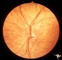
 |
H01 Panhypoplasia | Extreme hypoplasia. Very small disc. Peri-papillary halo (choroidal). Right eye. Note: normal vessels. Same patient as H_2. Anatomy: Optic disc. Pathology: Hypoplasia of the optic nerve. Disease/ Diagnosis: Hypoplasia. Clinical: Blind child. | Image |
| 2 |
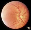
 |
H02 Panhypoplasia | Extreme hypoplasia. Very small disc. Peri-papillary halo (choroidal). Left eye. Note: normal vessels. Same patient as H_1. Anatomy: Optic disc. Pathology: Hypoplasia of the optic nerve. Disease/ Diagnosis: Hypoplasia. Clinical: Blind child. | Image |
| 3 |
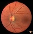
 |
H03 Panhypoplasia | Extreme hypoplasia. Note absence of retinal nerve fiber layer. Left eye. Girl. Same patient as H_4. Anatomy: Optic disc. Pathology: Hypoplasia of the optic nerve. Disease/ Diagnosis: Hypoplasia. Clinical: Left eye. Girl. | Image |
| 4 |
 |
H04 Panhypoplasia | Right eye. Normal eye. Girl. Same patient as H_3. Anatomy: Optic disc. Pathology: Hypoplasia of the optic nerve. Disease/ Diagnosis: Hypoplasia. | Image |
| 5 |
 |
H05 Panhypoplasia | Right eye. Distinctive septo-optic dysplasia.Hypoplasia of the optic nerve. Left eye normal. Amblyopic right eye. 24 year old woman. Anatomy: Optic disc. Pathology: Hypoplasia of the optic nerve. Disease/ Diagnosis: Hypoplasia. | Image |
| 6 |
 |
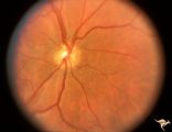
H06 Panhypoplasia | Bilateral hypoplasia. Top is Right eye - moderate. Bottom is Left eye - severe. Note venous tortuosity. Good example of double ring sign. De Morsier's syndrome.Septo-optic dysplasia. Same patient as H_7. Anatomy: Optic disc. Pathology: Hypoplasia of the optic nerve. Disease/ Diagnosis: Hypoplasia. ... | Image |
| 7 |
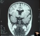 |
H07 Panhypoplasia | MRI Scan, coronal view showing absence of septum pellucidum. Hypoplastic chiasm. De Morsier's syndrome. Same patient as H_6. Anatomy: Optic disc. Pathology: Hypoplasia of the optic nerve. Disease/ Diagnosis: Hypoplasia. Imaging: MRI scan. | Image |
| 8 |
 |
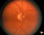
H08 Panhypoplasia | Severe hypoplasia. Right eye. Boy. Good example of double ring sign. Anatomy: Optic disc. Pathology: Hypoplasia of the optic nerve. Disease/ Diagnosis: Hypoplasia. | Image |
| 9 |
 |
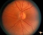
H09 Panhypoplasia | Moderate hypoplasia. Man. Anatomy: Optic disc. Pathology: Hypoplasia of the optic nerve. Disease/ Diagnosis: Hypoplasia. | Image |
| 10 |
 |
H10 Panhypoplasia | Cruzon's Disease. 26 year old man. Right eye. Mild hypoplasia. Son of patient in H_11 and H_12. Same patient in H_31. Father of patient in H_32. Anatomy: Optic disc. Pathology: Hypoplasia of the optic nerve. Disease/ Diagnosis: Hypoplasia. | Image |
| 11 |
 |
H11 Panhypoplasia | Cruzon's Disease. 47 year old woman. Right eye. Mild hypoplasia. Mother of patient in H_10 and H_31. Same patient as H_12. Grandmother of patient in H_32. Anatomy: Optic disc. Pathology: Hypoplasia of the optic nerve. Disease/ Diagnosis: Hypoplasia. | Image |
| 12 |
 |
H12 Panhypoplasia | Cruzon's Disease. 47 year old woman. Left eye. Mild hypoplasia. Mother of patient in H_10 and H_31. Same patient as H_11. Grandmother of patient in H_32. Anatomy: Optic disc. Pathology: Hypoplasia of the optic nerve. Pathology: Hypoplasia of the optic nerve. Disease/ Diagnosis: Hypoplasia. | Image |
| 13 |
 |
H13 Panhypoplasia | Right eye. Blind baby. Severe hypoplasia with blond fundus. Same patient as H_14. Anatomy: Optic disc. Pathology: Hypoplasia of the optic nerve. Disease/ Diagnosis: Hypoplasia. Imaging: Hypoplasia of the optic nerve. | Image |
| 14 |
 |
H14 Panhypoplasia | Left eye. Blind baby. Severe hypoplasia with blond fundus. Same patient as H_13. Anatomy: Optic disc. Pathology: Hypoplasia of the optic nerve. Disease/ Diagnosis: Hypoplasia. | Image |
| 15 |
 |
H15 Panhypoplasia | Moderate hypoplasia. Right eye. 14 year old boy. Good example of double ring sign. Same patient as H_16. Anatomy: Optic disc. Pathology: Hypoplasia of the optic nerve. Disease/ Diagnosis: Hypoplasia. | Image |
| 16 |
 |
H16 Panhypoplasia | Moderate hypoplasia. Left eye. 14 year old boy. Good example of double ring sign. Same patient as H_15. Anatomy: Optic disc. Pathology: Hypoplasia of the optic nerve. Disease/ Diagnosis: Hypoplasia. | Image |
| 17 |
 |
H17 Panhypoplasia | Bilateral mild hypoplasia without field defect. Right eye. 30 year old woman. Same patient as H_18. Anatomy: Optic disc. Pathology: Hypoplasia of the optic nerve. Disease/ Diagnosis: Hypoplasia. | Image |
| 18 |
 |
H18 Panhypoplasia | Bilateral mild hypoplasia without field defect. Left eye. 30 year old woman. Same patient as H_17. Anatomy: Optic disc. Pathology: Hypoplasia of the optic nerve. Disease/ Diagnosis: Hypoplasia. | Image |
| 19 |
 |
H19 Panhypoplasia | Mild hypoplasia with dysplasia in right eye. Right eye. Normal left eye. Same patient as H_20. Anatomy: Optic disc. Pathology: Hypoplasia of the optic nerve. Disease/ Diagnosis: Hypoplasia. | Image |
| 20 |
 |
H20 Panhypoplasia | Mild hypoplasia with dysplasia in right eye. Left eye. Same patient as H_19. Anatomy: Optic disc. Pathology: Hypoplasia of the optic nerve. Disease/ Diagnosis: Hypoplasia. | Image |
| 21 |
 |
H21 Panhypoplasia | Right eye. Hypoplasia with glial tissue haze. Same patient as H_22. Anatomy: Optic disc. Pathology: Hypoplasia of the optic nerve. Disease/ Diagnosis: Hypoplasia. | Image |
| 22 |
 |
H22 Panhypoplasia | Left eye. Normal disc. Same patient as H_21. Anatomy: Optic disc. Pathology: Hypoplasia of the optic nerve. Disease/ Diagnosis: Hypoplasia. | Image |
1 - 25 of 22