Best known for his world-renowned neuro-ophthalmology unit based at the University of California, San Francisco, William Hoyt, MD collected here more than 850 of his best images covering a wide range of disorders.
William F. Hoyt, MD, Professor Emeritus of Ophthalmology, Neurology and Neurosurgery, Department of Ophthalmology, University of California, San Francisco.
NOVEL: https://novel.utah.edu/
Filters: Collection: ehsl_novel_wfh
1 - 25 of 10
| Title | Description | Type | ||
|---|---|---|---|---|
| 1 |
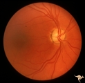 |
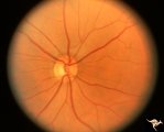
IF201a Temporal Cupping with Dominant Hereditary Optic Atrophy | 1969. Dominant hereditary optic atrophy (Kjer) Pair with IF2_1b. Right eye. Boy with reduced central acuity since childhood. Discs are pale temporally and the temporal nerve fiber layer is thin. Anatomy: Optic disc. Pathology: Dominant hereditary optic atrophy. Disease/ Diagnosis: Dominant hereditar... | Image |
| 2 |
 |
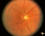
IF201b Temporal Cupping with Dominant Hereditary Optic Atrophy | 1969. Dominant hereditary optic atrophy (Kjer) Pair with IF2_1a. Left eye. Boy with reduced central acuity since childhood. and the temporal nerve fiber layer is thin. Anatomy: Optic disc. Pathology: Dominant hereditary optic atrophy. Disease/ Diagnosis: Dominant hereditary optic atrophy. Clinical: ... | Image |
| 3 |
 |
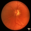
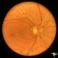
IF202a Temporal Cupping with Dominant Hereditary Optic Atrophy | Right eye. Teenage boy. Dominant hereditary optic atrophy (Kjer). Shows pallor and shallow cupping temporally. Pair with IF2_2b. 1975. Anatomy: Optic disc. Pathology: Dominant hereditary optic atrophy. Disease/ Diagnosis: Dominant hereditary optic atrophy. Clinical: Depressed central vision. | Image |
| 4 |
 |
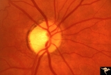
IF202b Temporal Cupping with Dominant Hereditary Optic Atrophy | Left eye. Teenage boy. Dominant hereditary optic atrophy (Kjer). Shows temporal pallor only. Shallow temporal cup. Pair with IF2_2a. 1975. Anatomy: Optic disc. Pathology: Dominant hereditary optic atrophy. Disease/ Diagnosis: Dominant hereditary optic atrophy. Clinical: Depressed central vision. | Image |
| 5 |
 |
IF203a Temporal Cupping with Dominant Hereditary Optic Atrophy | Right eye shows pallor and temporal cupping. Pair with IF2_3b. 1994. Anatomy: Optic disc. Pathology: Dominant hereditary optic atrophy. Disease/ Diagnosis: Dominant hereditary optic atrophy. Clinical: Depressed central vision. | Image |
| 6 |
 |
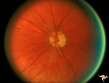
IF203b Temporal Cupping with Dominant Hereditary Optic Atrophy | Left disc is hypoplastic and smaller with temporal pallor. Pair with IF2_3a. 1994. Anatomy: Optic disc. Pathology: Dominant hereditary optic atrophy. Disease/ Diagnosis: Dominant hereditary optic atrophy. Clinical: Depressed central vision. | Image |
| 7 |
 |
IF204a Temporal Cupping with Dominant Hereditary Optic Atrophy | Right eye with temporal pallor and shallow cupping. Pair with IF2_4b. 1960. Anatomy: Optic disc. Pathology: Dominant hereditary optic atrophy. Disease/ Diagnosis: Dominant hereditary optic atrophy. Clinical: Dominant hereditary optic atrophy. | Image |
| 8 |
 |
IF204b Temporal Cupping with Dominant Hereditary Optic Atrophy | Left eye with temporal pallor and shallow cupping. Pair with IF2_4a. 1960. Anatomy: Optic disc. Pathology: Dominant hereditary optic atrophy. Disease/ Diagnosis: Dominant hereditary optic atrophy. Clinical: Depressed central vision. | Image |
| 9 |
 |
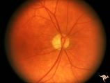
IF205a Temporal Cupping with Dominant Hereditary Optic Atrophy | 1970. Right eye. Pair with IF2_5b. 55 year old woman with deficient vision all her life. Typical pattern of dominant hereditary atrophy. Temporal pallor and shallow cupping. Anatomy: Optic disc. Pathology: Dominant hereditary optic atrophy. Disease/ Diagnosis: Dominant hereditary optic atrophy. Cli... | Image |
| 10 |
 |
IF205b Temporal Cupping with Dominant Hereditary Optic Atrophy | 1970. Left eye. Pair with IF2_5a. 55 year old woman with deficient vision all her life. Typical pattern of dominant hereditary atrophy. Temporal pallor and shallow cupping. Anatomy: Optic disc. Pathology: Dominant hereditary optic atrophy. Disease/ Diagnosis: Dominant hereditary optic atrophy. Clin... | Image |
1 - 25 of 10