Best known for his world-renowned neuro-ophthalmology unit based at the University of California, San Francisco, William Hoyt, MD collected here more than 850 of his best images covering a wide range of disorders.
William F. Hoyt, MD, Professor Emeritus of Ophthalmology, Neurology and Neurosurgery, Department of Ophthalmology, University of California, San Francisco.
NOVEL: https://novel.utah.edu/
Filters: Collection: ehsl_novel_wfh
1 - 25 of 16
| Title | Description | Type | ||
|---|---|---|---|---|
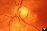
| 1 |
 |
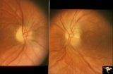
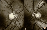
Segmental Atrophy - Hemianopic (Band) Atrophy | Segmental Atrophy - Band atrophy from right optic tract injury. Red free filter. Left eye. Has temporal hemianopia with band atrophy. Note loss of nasal nerve fiber layer. Old right optic tract injury. 1972. Pair with IIA2C_9a. Anatomy: Optic disc. Pathology: Right optic tract injury. Disease/Diagno... | Image |
| 2 |
 |
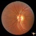
Segmental Atrophy - Hemianopic (Band) Atrophy | Segmental Atrophy - Band atrophy with horizontal cupping. Pituitary adenoma. Magnification of 14a. Pair with IIA2C_14a. 1975. Anatomy: Optic disc. Pathology: Chiasmal compression from pituitary adenoma in a cupped disc. Disease/Diagnosis: Band atrophy and cupping. Clinical: Temporal hemianopia. | Image |
| 3 |
 |
Segmental Atrophy - Hemianopic (Band) Atrophy | Segmental Atrophy - Band atrophy with horizontal cupping. Transverse cup. Pair with IIA2C_14b. 1975. Anatomy: Optic disc. Pathology: Chiasmal compression from pituitary adenoma in a cupped disc. Disease/Diagnosis: Band atrophy and cupping. Clinical: Temporal hemianopia. | Image |
| 4 |
 |
Segmental Atrophy - Hemianopic (Band) Atrophy | Segmental Atrophy - Band atrophy from right optic tract injury. Red free filter. Left eye. Has temporal hemianopia with band atrophy. Note loss of nasal nerve fiber layer. Old right optic tract injury. 1972. Pair with IIA2C_9b. Anatomy: Optic disc. Pathology: Right optic tract injury. Disease/Diagno... | Image |
| 5 |
 |
Segmental Atrophy - Hemianopic (Band) Atrophy | Segmental Atrophy - Band atrophy from right optic tract injury. This eye has a nasal hemianopia. Its disc shows temporal pallor with an intact nasal nerve fiber layer. Old right optic tract injury. 1986. Pair with IIA2C_8b. Anatomy: Optic disc. Pathology: Right optic tract injury. Disease/Diagnosis:... | Image |
| 6 |
 |
Segmental Atrophy - Hemianopic (Band) Atrophy | Segmental Atrophy - Band atrophy from right optic tract injury. Left eye. Has temporal hemianopia with band atrophy. Note loss of nasal nerve fiber layer. Old right optic tract injury. 1986. Pair with IIA2C_8a. Anatomy: Optic disc. Pathology: Right optic tract injury. Disease/Diagnosis: Homonymous h... | Image |
| 7 |
 |
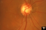
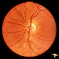
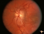
Segmental Atrophy - Hemianopic (Band) Atrophy | Segmental Atrophy - Band atrophy with papilledema. 1975. Patient had a right optic tract glioma. Anatomy: Optic disc. Pathology: Glioma of the right optic tract. Disease/Diagnosis: Twin peaks papilledema. Clinical: Left homonymous hemianopia. | Image |
| 8 |
 |
Segmental Atrophy - Hemianopic (Band) Atrophy | Segmental Atrophy - Magnification of IIA2C_02a. Band atrophy in an eye with temporal hemianopia. Wyburn-Mason Syndrome extending to the chiasm. Left eye. 1975. Right eye in patient was blind. Anatomy: Optic disc. Pathology: Right sided chiasmal AVM. Disease/Diagnosis: Band atrophy due to chiasmal A... | Image |
| 9 |
 |
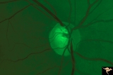
Segmental Atrophy - Hemianopic (Band) Atrophy | Segmental Atrophy - Hemianopic (band) atrophy - Bilateral horizontal band atrophy secondary to old chiasmal trauma. Note the presence of arcuate nerve fibers and the absence of temporal and nasal nerve fibers. Note the sharp edged pallor of the nasal disc margin. Right eye. Pair with IIA2C_1b. 1985.... | Image |
| 10 |
 |
Segmental Atrophy - Hemianopic (Band) Atrophy | Segmental Atrophy - Hemianopic (band) atrophy - Bilateral horizontal band atrophy secondary to old chiasmal trauma. Note the presence of arcuate nerve fibers and the absence of temporal and nasal nerve fibers. Note the sharp edged pallor of the nasal disc margin. Right eye. Pair with IIA2C_1a. 1985.... | Image |
| 11 |
 |
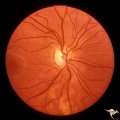
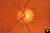
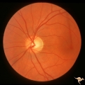
Segmental Atrophy - Hemianopic (Band) Atrophy | Segmental Atrophy - Band atrophy. Shows band atrophy in left disc with preserved upper and lower arcuate nerve fiber bundles. Right disc has thinning of both upper and lower arcuate nerve fiber bundles, temporal pallor, and an intact nasal nerve fiber layer. 1972. Anatomy: Optic disc. Pathology: Rig... | Image |
| 12 |
 |
Segmental Atrophy - Hemianopic (Band) Atrophy | Segmental Atrophy - Band atrophy with ""Twin Peaks"" papilledema. Central band of the optic disc is completely atrophic and does not swell. ""Axons that are not there can not swell."" Anatomy: Optic disc. Pathology: Optic tract injury. Disease/Diagnosis: Twin peaks papilledema. Clinical: Left homony... | Image |
| 13 |
 |
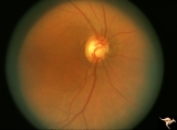
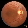
Segmental Atrophy - Hemianopic (Band) Atrophy | Segmental Atrophy - Band atrophy with temporal hemianopia. 1983. Anatomy: Optic disc. Pathology: Atrophy of the chiasm or left optic tract. Disease/Diagnosis: Segmental band atrophy. Clinical: Right temporal field defect. | Image |
| 14 |
 |
Segmental Atrophy - Hemianopic (Band) Atrophy | Segmental Atrophy - Band atrophy in an eye with temporal hemianopia. Wyburn-Mason Syndrome extending to the chiasm. Left eye 1975. Anatomy: Optic disc. Pathology: Right sided chiasmal AVM. Disease/Diagnosis: Band atrophy due to chiasmal AVM and Wyburn-Mason Syndrome. Clinical: Blind right eye, temp... | Image |
| 15 |
 |
Segmental Atrophy - Hemianopic (Band) Atrophy from Eight Optic Tract Injury | Segmental Atrophy - Band atrophy from right optic tract injury. This eye has a nasal hemianopia. Its disc shows temporal pallor with an intact nasal nerve fiber layer. Pair with IIA2C_7b. Anatomy: Optic disc. Pathology: Right optic tract injury. Disease/Diagnosis: Homonymous hemioptic atrophy. Clini... | Image |
| 16 |
 |
Segmental Atrophy - Hemianopic (Band) Atrophy from Eight Optic Tract Injury | Segmental Atrophy - Band atrophy from right optic tract injury. Left eye. Has temporal hemianopia with band atrophy. Note loss of nasal nerve fiber layer. Four and a half months after injury from intracranial pressure catheter. Pair with IIA2C_7a. Anatomy: Optic disc. Pathology: Right optic tract in... | Image |
1 - 25 of 16