Best known for his world-renowned neuro-ophthalmology unit based at the University of California, San Francisco, William Hoyt, MD collected here more than 850 of his best images covering a wide range of disorders.
William F. Hoyt, MD, Professor Emeritus of Ophthalmology, Neurology and Neurosurgery, Department of Ophthalmology, University of California, San Francisco.
NOVEL: https://novel.utah.edu/
TO
Filters: Collection: ehsl_novel_wfh
| Title | Description | Type | ||
|---|---|---|---|---|
| 1 |
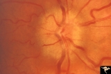 |
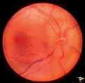
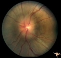
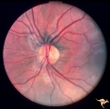
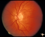
Unilateral Papilledema | Unilateral papilledema due to elevated intracranial pressure. Right eye. This eye has papilledema. Patient described transient monocular blindness with turning right eye. Asymmetric papilledema. Anatomy: Optic disc. Pathology: Unilateral papilledema. Disease/Diagnosis: Iidiopathic intracranial hyper... | Image |
| 2 |
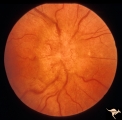 |
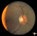
Unilateral Papilledema | Right eye.Chronic Papilledema in right eye. Woman. Pseudo Foster Kennedy due to asymmetric papilledema. Anatomy: Optic disc. Pathology: Chronic papilledema; optic atrophy. Disease/Diagnosis: Idiopathic intracranial hypertension (pseudotumor cerebri) causing pseudo Foster-Kennedy Syndrome. Clinical: ... | Image |
| 3 |
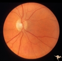 |
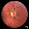
Unilateral Papilledema | Right eye. Atrophic nerve right eye. Large falx meningioma. True Foster Kennedy Syndrome. Anatomy: Optic disc. Pathology: Chronic papilledema; optic atrophy. Disease/Diagnosis: Meningioma causing Foster-Kennedy Syndrome. Clinical: Visual loss one eye; Transient visual obscuration other eye. | Image |
| 4 |
 |
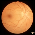
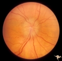
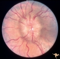
Unilateral Papilledema | Left eye. Has no optic cup. 24 year old obese woman. Anatomy: Optic disc. Pathology: Unilateral papilledema. Disease/Diagnosis: Idiopthatic intracranial hypertension, pseudotumor cerebri. Clinical: Woman, headache, transient visual obscurations. | Image |
| 5 |
 |
Unilateral Papilledema | Left eye. Left eye has papilledema. Large falx meningioma. True Foster Kennedy Syndrome. Anatomy: Optic disc. Pathology: Chronic papilledema; optic atrophy. Disease/Diagnosis: Meningioma causing Foster-Kennedy Syndrome. Clinical: Visual loss one eye; transient visual obscuration other eye. | Image |
| 6 |
 |
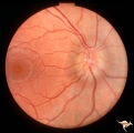
Unilateral Papilledema | Right eye. Mild disc blurring. Obese woman complaining of headaches. Asymmetric papilledema. Anatomy: Optic disc. Pathology: Asymmetric papilledema. Disease/Diagnosis: Idiopathic intracranial hypertension, pseudotumor cerebri. Clinical: Headache, obese woman. | Image |
| 7 |
 |
Unilateral papilledema | Unilateral papilledema in Pseudotumor cerebri in patient with elevated intracranial pressure. Right eye. Anatomy: Optic disc. Pathology: Unilateral papilledema. Disease/Diagnosis: Idiopathic intracranial hypertension (pseudotumor cerebri). Clinical: Transient monocular blindness (transient visual ob... | Image |
| 8 |
 |
Unilateral Papilledema | Unilateral papilledema in Pseudotumor cerebri in patient with elevated intracranial pressure. Right eye. Anatomy: Optic disc. Pathology: Unilateral papilledema. Disease/Diagnosis: Idiopathic intracranial hypertension (pseudotumor cerebri). Clinical: Transient monocular blindness (transient visual ob... | Image |
| 9 |
 |
Unilateral Papilledema | Unilateral papilledema in Pseudotumor cerebri in patient with elevated intracranial pressure. Left eye. Has no optic cup. Disc is flat. Anatomy: Optic disc. Pathology: Unilateral papilledema. Disease/Diagnosis: Idiopathic intracranial hypertension (pseudotumor cerebri). Clinical: Transient monocular... | Image |
| 10 |
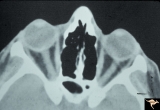 |
Unilateral Papilledema | CT scan showing equal thickening of the optic nerves. CT scan of no help in determining which eye has the papilledema. In this case, right eye has papilledema. Scan of patient depicted P_13a and P_13b. Anatomy: Optic disc. Pathology: Unilateral papilledema. Disease/Diagnosis: Idiopathic intracranial... | Image |
| 11 |
 |
Unilateral Papilledema | Left eye. Patient had tumor on right side. Right sided large meningioma. optociliary shunt at 10:00. Foster Kennedy. Anatomy: Optic disc. Pathology: Unilateral papilledema. Disease/Diagnosis: Meningioma of the brain. Clinical: Headache. | Image |
| 12 |
 |
Unilateral Papilledema | Right eye. Woman. Anomalous optic disc elevation in right eye only. This woman's case mimics unilateral papilledema from pseudotumor cerebri. Anatomy: Optic disc. Pathology: Anomalous disc elevation. Disease/Diagnosis: Pseudo papilledema. Clinincal: Mimic of papilledema; symptoms: headache; signs: a... | Image |
| 13 |
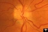 |
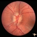
Unilateral Papilledema | Unilateral papilledema due to intracranial pressure. Left eye. This eye shows minor disc blur, inferiorly. No optic cup. Disc is flat. Anatomy: Optic disc. Pathology: Unilateral papilledema. Disease/Diagnosis: Idiopathic intracranial hypertension (pseudotumor cerebri). Clinical: Gaze evoked blindnes... | Image |
| 14 |
 |
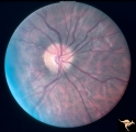
Unilateral Papilledema | Right eye. Has papilledema. Patient has pseudotumor cerebri. 24 year old obese woman. Anatomy: Optic disc. Pathology: Unilateral papilledema. Disease/Diagnosis: Idiopathic intracranial hypertension, pseudotumor cerebri. Clinical: Woman, headache, transient visual obscurations. | Image |
| 15 |
 |
Unilateral Papilledema | Left eye. Has no optic cup. Woman. Left optic disc is flat and cupless. This woman's case mimics unilateral papilledema from pseudotumor cerebri. Anatomy: Optic disc. Pathology: Anomalous disc elevation. Disease/Diagnosis: Pseudo papilledema. Clinical: Mimic of papilledema; symptoms: headache; signs... | Image |
| 16 |
 |
Unilateral Papilledema | Left eye. Low grade papilledema. Obese woman complaining of headaches. Asymmetric papilledema. Anatomy: Optic disc. Pathology: Asymmetric papilledema. Disease/Diagnosis: Idiopathic intracranial hypertension, pseudotumor cerebri. Clinical: Headache, obese woman. | Image |
| 17 |
 |
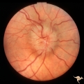
Unilateral Papilledema | Right eye. Has slight disc blur. Asymmetric papilledema. 35 year old woman. Anatomy: Optic disc. Pathology: Unilateral papilledema. Disease/Diagnosis: Idiopathic intracranial hypertension (pseudotumor cerebri). Clinical: Gaze evoked blindness. | Image |
| 18 |
 |
Unilateral Papilledema | Left eye. This eye has papilledema. 35 year old woman. Anatomy: Optic disc. Pathology: Unilateral papilledema. Disease/Diagnosis: Idiopathic intracranial hypertension (pseudotumor cerebri). Clinical: Gaze evoked blindness. | Image |
| 19 |
 |
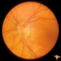
Unilateral Papilledema | Left eye. Has papilledema. 27 year old white woman. Anatomy: Optic disc. Pathology: Unilateral papilledema. Disease/Diagnosis: Idiopathic intracranial hypertension, pseudotumor cerebri. Clinical: Woman, headache. | Image |
| 20 |
 |
Unilateral Papilledema | Left eye. Has papilledema. Patient has Pseudotumor cerebri. Woman. Anatomy: Optic disc. Pathology: Unilateral papilledema. Disease/Diagnosis: Idiopathic intracranial hypertension, pseudotumor cerebri. Clinical: Woman, headache, transient visual obscuration. | Image |
| 21 |
 |
Unilateral Papilledema | Unilateral papilledema in pseudotumor cerebri. Right eye. Has no cup. 27 year old white woman. Anatomy: Optic disc. Pathology: Unilateral papilledema. Disease/Diagnosis: Idiopathic intracranial hypertiension, pseudotumor cerebri. Clinical: Woman, headache. | Image |
| 22 |
 |
Unilateral Papilledema | Left eye. Flat cupless disc. Woman. Anatomy: Optic disc. Pathology: Chronic papilledema; optic atrophy. Disease/Diagnosis: Idiopathic intracranial hypertension (pseudotumor cerebri) causing pseudo Foster-Kennedy Syndrome. Clinical: Visual loss in atrophic eye; obese. | Image |
| 23 |
 |
Unilateral Papilledema | Unilateral papilledema in Pseudotumor cerebri in patient with elevated intracranial pressure. Left eye. Has no optic cup. Disc is flat. Anatomy: Optic disc. Pathology: Unliateral papilledema. Disease/Diagnosis: Idiopathic intracranial hypertension (pseudotumor cerbri). Clinical: Transient monocular ... | Image |
| 24 |
 |
Unilateral Papilledema | Right eye. Patient had tumor on right side. Right sided large meningioma. Disc edema due to tumor. 29 year old black woman. The right disc has mild temporal pallor. Anatomy: Optic disc. Pathology: Uninaleral papilledema. Disease/Diagnosis: Meningioma of the brain. Clinical: Headache. | Image |
| 25 |
 |
Unilateral Papilledema | Right eye. Has no optic cup. Patient has pseudotumor cerebri. Woman. Anatomy: Optic disc. Pathology: Unilateral papilledema. Disease/Diagnosis: Idiopathic intracranial hypertension, pseudotumor cerebri. Clinical: Woman, headache, transient visual obscurations. | Image |