Best known for his world-renowned neuro-ophthalmology unit based at the University of California, San Francisco, William Hoyt, MD collected here more than 850 of his best images covering a wide range of disorders.
William F. Hoyt, MD, Professor Emeritus of Ophthalmology, Neurology and Neurosurgery, Department of Ophthalmology, University of California, San Francisco.
NOVEL: https://novel.utah.edu/
TO
Filters: Collection: ehsl_novel_wfh
1 - 25 of 4
| Title | Description | Type | ||
|---|---|---|---|---|
| 1 |
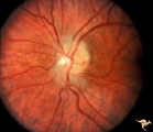 |
F202 Optic Nerve Sheath Meningioma | Optic nerve sheath meningioma. Note optociliary vein at 3:00. The disc is atrophic. Anatomy: Optic disc. Pathology: Chronic optic disc swelling caused by optic nerve sheath meningioma. Disease/ Diagnosis: Chronic optic disc swelling caused by optic nerve sheath meningioma. | Image |
| 2 |
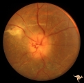 |
F203 Optic Nerve Sheath Meningioma | Optic nerve sheath meningioma. Note optociliary vessels on the disc. The disc is partially atrophic and blurred by previous edema. The cause of the choroidal scar was not determined. Anatomy: Optic disc. Pathology: Chronic optic disc swelling caused by optic nerve sheath meningioma. DIsease/ Diagnos... | Image |
| 3 |
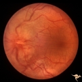 |
F204 Optic Nerve Sheath Meningioma | Optic disc swelling due to meningioma. Notice choroidal folds through the macula of left eye. Anatomy: Optic disc. Pathology: Chronic optic disc swelling caused by optic nerve sheath meningioma. Disease/ Diagnosis: Chronic optic disc swelling caused by optic nerve sheath meningioma. | Image |
| 4 |
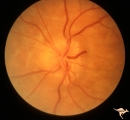 |
F201 Optic Nerve Sheath Meningioma | Right eye. Woman with ophthalmoplegia proptosis for 14 years. Visual field reduced due to optic nerve sheath meningioma. Notice large optociliary vessel temporally. Anatomy: Optic disc. Pathology: Chronic optic disc swelling caused by optic nerve sheath meningioma. Disease/ Diagnosis: Chronic optic ... | Image |
1 - 25 of 4