Best known for his world-renowned neuro-ophthalmology unit based at the University of California, San Francisco, William Hoyt, MD collected here more than 850 of his best images covering a wide range of disorders.
William F. Hoyt, MD, Professor Emeritus of Ophthalmology, Neurology and Neurosurgery, Department of Ophthalmology, University of California, San Francisco.
NOVEL: https://novel.utah.edu/
TO
Filters: Collection: ehsl_novel_wfh
1 - 25 of 13
| Title | Description | Type | ||
|---|---|---|---|---|
| 1 |
 |
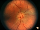
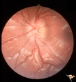
F2b01 Optic Nerve Glioma | Left eye. Woman with optic nerve glioma. Anatomy: Optic disc. Pathology: Optic nerve swelling secondary to retrobulbar optic glioma. Disease/ Diagnosis: Optic nerve glioma. | Image |
| 2 |
 |
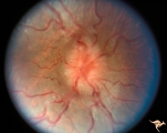
F2b05 Optic Disc Swelling from Optic Glioma | Optic disc swelling from optic glioma. Patient had Neurofibromatosis (NF1). Left eye. 7 year old girl. 20/100 acuity. Glioma of the left optic nerve. Anatomy: Optic disc. Pathology: Optic nerve glioma. Disease/ Diagnosis: Optic nerve swelling secondary to retrobulbar optic glioma | Image |
| 3 |
 |
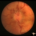
F2b06 Optic Disc Swelling from Optic Glioma | Right eye. Optic glioma with disc swelling. Anatomy: Optic disc. Pathology: Optic nerve glioma. Disease/ Diagnosis: Optic nerve swelling secondary to retrobulbar optic glioma. | Image |
| 4 |
 |
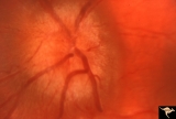
F2b07 Optic Disc Swelling from Optic Glioma | 6 year old with neurofibromatosis (NF1). Right eye went blind. Light perception. Optic canal enlargement due to glioma. Notice optociliary vessels. Same patient as F2b_8. Anatomy: Optic disc. Pathology: Optic nerve glioma. Disease/ Diagnosis: Optic nerve swelling secondary to retrobulbar optic gliom... | Image |
| 5 |
 |
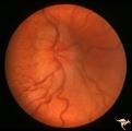
F2b08 Optic Disc Swelling from Optic Glioma | Left eye. Optic nerve glioma. Disc swelling without visual loss. Same patient as F2b_7. Anatomy: Optic disc. Pathology: Optic nerve glioma. Disease/ Diagnosis: Optic nerve swelling secondary to retrobulbar optic glioma. | Image |
| 6 |
 |
F2b11 Optic Disc Swelling from Optic Glioma | Optic disc swelling from optic glioma. Note the signs of vein occlusion and the optociliary bypass vien at 4:00. Left eye. Anatomy: Optic disc. Pathology: Optic nerve glioma. Disease/ Diagnosis: Optic nerve swelling secondary to retrobulbar optic glioma. | Image |
| 7 |
 |
F2b13 Progression of Optic Disc Changes Caused by Malignant Optic Nerve Glioma of Adulthood | Progression. Group with F2b_12_1 and F2b_14_3. 69 year old male. April 22, 1992. There are signs of CVRO. Reference: Hoyt WF, Meshel LG, Lessell S, Schatz NJ, Suckling RD. Malignant optic glioma of adulthood. Brain. 1973;96(1):121-32. Anatomy: Optic disc. Pathology: Optic nerve glioma. Disease/ Diag... | Image |
| 8 |
 |
F2b14 Progression of Optic Disc Changes Caused by Malignant Optic Nerve Glioma of Adulthood | Progression. Group with F2b_12_1 and F2b_13_2. 69 year old male. Shows signs of myelin being squeezed through the optic disc into the eye. June 6, 1992. Reference: Hoyt WF, Meshel LG, Lessell S, Schatz NJ, Suckling RD. Malignant optic glioma of adulthood. Brain. 1973;96(1):121-32. Anatomy: Optic di... | Image |
| 9 |
 |
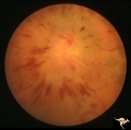
F2b02 Progressive Optic Disc Swelling with Optic Glioma | Progressive optic disc swelling with optic glioma. Left eye. Woman with optic disc swelling. April 1969. Same patient as F2b_03 and F2b_04. Anatomy: Optic disc. Pathology: Optic nerve swelling secondary to retrobulbar optic glioma. Disease/ Diagnosis: Optic nerve glioma. | Image |
| 10 |
 |
F2b03 Progressive Optic Disc Swelling with Optic Glioma | Progressive optic disc swelling with optic glioma. Left eye. Woman with optic disc swelling. Edema is becoming pale. May 1969. Same patient as F2b_02 and F2b_04. Anatomy: Optic disc. Pathology: Optic nerve swelling secondary to retrobulbar optic glioma. Disease/ Diagnosis: Optic nerve glioma. | Image |
| 11 |
 |
F2b04 Progressive Optic Disc Swelling with Optic Glioma | Progressive optic disc swelling with optic glioma. Left eye. Woman with optic disc swelling. Entire disc obscured by overlying edema and hemorrhage. Blind in 3 months. This series illustrates a progressive infarction of the optic disc adjacent to an optic disc glioma. June 1969. Same patient as F2b_... | Image |
| 12 |
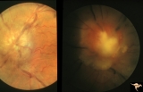 |
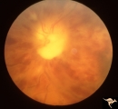
F2b09 Optic Disc Swelling from Malignant Optic Nerve Glioma | Malignant optic nerve glioma of adulthood with blindness and optic disc edema. Right image shows white material extruded from the swollen optic disc. This material is myelin being squeezed into the eye from the nerve infarction. Autopsy specimen of this eye shown in F2b_10. Reference: Hoyt WF, Meshe... | Image |
| 13 |
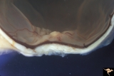 |
F2b10 Malignant Optic Nerve Glioma, Gross Pathologic Specimen | Pathologic specimen of optic nerve glioma shown in slide F2b_09. White material on top of swollen disc is myelin. Reference: Hoyt WF, Meshel LG, Lessell S, Schatz NJ, Suckling RD. Malignant optic glioma of adulthood. Brain. 1973;96(1):121-32. Anatomy: Optic disc. Pathology: Optic nerve glioma. Disea... | Image |
1 - 25 of 13