Best known for his world-renowned neuro-ophthalmology unit based at the University of California, San Francisco, William Hoyt, MD collected here more than 850 of his best images covering a wide range of disorders.
William F. Hoyt, MD, Professor Emeritus of Ophthalmology, Neurology and Neurosurgery, Department of Ophthalmology, University of California, San Francisco.
NOVEL: https://novel.utah.edu/
Filters: Collection: ehsl_novel_wfh
1 - 25 of 10
| Title | Description | Type | ||
|---|---|---|---|---|
| 1 |
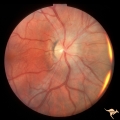 |
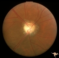
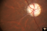
ID01 Post Papilledema Gliosis | Post papilledema milky gliosis with arteriolar constriction, 1982, right eye, pair with ID_2. Anatomy: Optic disc. Pathology: Post papilledema atrophy and gliosis due to huge anterior communicating artery aneurysm. Disease/ Diagnosis: Elevated intracranial pressure from aneurysm. Clinical: Diminishe... | Image |
| 2 |
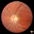 |
ID02 Post Papilledema Gliosis | Post papilledema milky gliosis with arteriolar constriction and atrophy, 1982, left eye, pair with ID_1. Anatomy: Optic disc. Pathology: Post papilledema atrophy and gliosis due to huge anterior communicating artery aneurysm. Disease/ Diagnosis: Elevated intracranial pressure from aneurysm. Clinical... | Image |
| 3 |
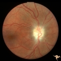 |
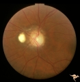
ID03a Post Papilledema Atrophy with Marked Gliosis | Post papilledema atrophy with marked gliosis in a patient with pseudotumor cerebri. Patient weighed over 300 pounds. Right eye blind. 1981. Right eye. Pair with ID_3b. Anatomy: Optic disc. Pathology: Post papilledema atrophy and gliosis from long standing elevated intracranial pressure. Disease/ Dia... | Image |
| 4 |
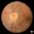 |
ID03b Post Papilledema Atrophy with Marked Gliosis | Post papilledema atrophy with marked gliosis in a patient with pseudotumor cerebri. Patient weighed over 300 pounds. Left eye has visual field defects. 1981, right eye, pair with ID_3a. Anatomy: Optic disc. Pathology: Post papilledema atrophy and gliosis from long standing elevated intracranial pres... | Image |
| 5 |
 |
ID04a Post Papilledema Atrophy with Marked Gliosis | Post papilledema atrophy with marked gliosis in a patient with pseudotumor cerebri, 1985, right eye, pair with ID_4b, Note "high water" marks in peripapillary pigment epithelial layer. Anatomy: Optic disc. Pathology: Post papilledema atrophy and gliosis from long standing elevated intracranial press... | Image |
| 6 |
 |
ID04b Post Papilledema Atrophy with Marked Gliosis | Post papilledema atrophy with marked gliosis in a patient with pseudotumor. Nasal ovoid absence of the retinal pigment epithelium. Presumably a defect from the long standing papilledema. 1985,. Right eye, pair with ID_4a. Anatomy: Optic disc. Pathology: Post papilledema atrophy and gliosis from long... | Image |
| 7 |
 |
ID05a Post Papilledema Optic Atrophy from Pseudotumor Cerebri | Left eye, October 1999, Post papilledema optic atrophy from pseudotumor cerebri. Note optociliary veins in both discs. Gliosis and partial pallor following long standing papilledema and intracranial pressure. Anatomy: Optic disc. Pathology: Post papilledema atrophy and gliosis from long standing el... | Image |
| 8 |
 |
ID05b Post Papilledema Optic Atrophy from Pseudotumor Cerebri | Right eye, October 1999, Post papilledema optic atrophy from pseudotumor cerebri. Note optociliary veins in both discs. Gliosis and partial pallor following long standing papilledema and intracranial pressure. Anatomy: Optic disc. Pathology: Post papilledema atrophy and gliosis from long standing el... | Image |
| 9 |
 |
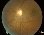
ID06 Post Papilledema Optic Atrophy with Arteriolar Sheathing and Optociliary Veins | 1989. Post papilledema optic atrophy with arteriolar sheathing and optociliary veins. Anatomy: Optic disc. Pathology: Long standing effects of intracranial pressure. Clinical: Blindness. | Image |
| 10 |
 |
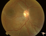
ID07 Post Papilledema Optic Atrophy | Post papilledema optic atrophy with gliosis and arteriolar narrowing. 1994. Anatomy: Optic disc. Pathology: Residue of long standing papilledema. Clinical: Visual loss. | Image |
1 - 25 of 10