Best known for his world-renowned neuro-ophthalmology unit based at the University of California, San Francisco, William Hoyt, MD collected here more than 850 of his best images covering a wide range of disorders.
William F. Hoyt, MD, Professor Emeritus of Ophthalmology, Neurology and Neurosurgery, Department of Ophthalmology, University of California, San Francisco.
NOVEL: https://novel.utah.edu/
TO
Filters: Collection: ehsl_novel_wfh
1 - 25 of 15
| Title | Description | Type | ||
|---|---|---|---|---|
| 1 |
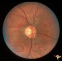 |
H101 Occipital Hemianoptic Hypoplasia | Right eye. Same patient as H_102. Anatomy: Optic disc. Pathology: Occipital hemianoptic hypoplasia. Disease/ Diagnosis: Congenital defect of the occipital lobe. | Image |
| 2 |
 |
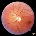
H102 Occipital Hemianoptic Hypoplasia | Left eye. Trans-synaptic band atrophy. Left homonymous hemianopia from right occipital porencephaly. Loss of nasal nerve fibers. Same patient as H_101. Anatomy: Optic disc. Pathology: Occipital hemianoptic hypoplasia. Disease/ Diagnosis: Congenital defect of the occipital lobe. | Image |
| 3 |
 |
H103 Occipital Hemianoptic Hypoplasia | Right eye. Congenital right homonymous hemianopia. Absent nerve fiber layer in right eye. Same patient as H_104. Anatomy: Optic disc. Pathology: Occipital hemianoptic hypoplasia. Disease/ Diagnosis: Congenital defect of the occipital lobe. | Image |
| 4 |
 |
H104 Occipital Hemianoptic Hypoplasia | Left eye. Contrast with nasal nerve fiber in right eye, H_103. Anatomy: Optic disc. Pathology: Occipital hemianoptic hypoplasia. Disease/ Diagnosis: Congenital defect of the occipital lobe. | Image |
| 5 |
 |
H105 Occipital Hemianoptic Hypoplasia | Left congenital homonymous hemianopia. Right occipital AVM. Nasal nerve fiber layer loss in left eye. Compare with right eye. Same patient as H_106. Anatomy: Optic disc. Pathology: Occipital hemianoptic hypoplasia. DIsease/ Diagnosis: Congenital defect of the occipital lobe | Image |
| 6 |
 |
H106 Occipital Hemianoptic Hypoplasia | Same patient as H_105. Anatomy: Optic disc. Pathology: Occipital hemianoptic hypoplasia. Disease/ Diagnosis: Congenital defect of the occipital lobe. | Image |
| 7 |
 |
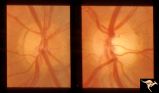
H89 Occipital Hemianoptic Hypoplasia | Diagram of homonymous hemioptic hypoplasia showing pattern of preserved nerve fibers. Homonymous hemioptic hypoplasia. Fundoscopic features in standard and red-free illumination in three patients with congenital hemiplegia. Anatomy: Optic disc. Pathology: Occipital hemianoptic hypoplasia. Disease/ D... | Image |
| 8 |
 |
H90 Occipital Hemianoptic Hypoplasia | Note left disc (right side of image) is the eye with temporal field defect. Shows band atrophy. Anatomy: Optic disc. Pathology: Occipital hemianoptic hypoplasia. Congenital defect of the occipital lobe. | Image |
| 9 |
 |
H91 Occipital Hemianoptic Hypoplasia | Left eye with temporal field defect shows trans-synaptic band atrophy. Same patient as H_92. Anatomy: Optic disc. Pathology: Occipital hemianoptic hypoplasia. Disease/ Diagnosis: Congenital defect of the occipital lobe. | Image |
| 10 |
 |
H92 Occipital Hemianoptic Hypoplasia | Right eye. Same patient as H_91. Anatomy: Optic disc. Pathology: Occipital hemianoptic hypoplasia. Disease/ Diagnosis: Congenital defect of the occipital lobe. | Image |
| 11 |
 |
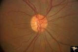
H93 Occipital Hemianoptic Hypoplasia | Visual field. Left eye. Right inferior homonymous. Same patient as H_94, H_95, H_96, H_97. Anatomy: Optic disc. Pathology: Occipital hemianoptic hypoplasia. Disease/ Diagnosis: Congenital defect of the occipital lobe. Imaging: MRI scan - See slide H97. | Image |
| 12 |
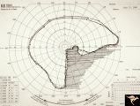 |
H94 Occipital Hemianoptic Hypoplasia | Visual field. Right eye. Quatrantanopia. Same patient as H_93, H_95, H_96, H_97. Anatomy: Optic disc. Pathology: Occipital hemianoptic hypoplasia. Disease/ Diagnosis: Congenital defect of the occipital lobe. Imaging: MRI scan - See slide H97. | Image |
| 13 |
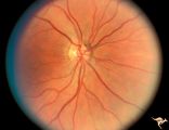 |
H95 Occipital Hemianoptic Hypoplasia | Right eye with temporal field defect shows trans-synaptic band atrophy. Absence of nasal nerve fibers. Same patient as H_93, H_94, H_96, H_97. Anatomy: Optic disc. Pathology: Occipital hemianoptic hypoplasia. Disease/ Diagnosis: Congenital defect of the occipital lobe. Imaging: MRI scan - See slide ... | Image |
| 14 |
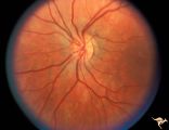 |
H96 Occipital Hemianoptic Hypoplasia | Left eye has nasal quadrantic field defect. Same patient as H_93, H_94, H_95, H_97. Anatomy: Optic disc. Pathology: Occipital hemianoptic hypoplasia. Disease/ Diagnosis: Congenital defect of the occipital lobe. Imaging: MRI scan - See slide H97. | Image |
| 15 |
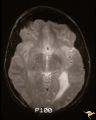 |
H97 Occipital Hemianoptic Hypoplasia | MRI scan shows left occipital lobe periventricular leuko-melacia. Same patient as H_93, H_94, H_95, H_96. Anatomy: Optic disc. Pathology: Occipital hemianoptic hypoplasia. DIsease/ Diagnosis: Congenital defect of the occipital lobe. Imaging: MRI scan. | Image |
1 - 25 of 15