Best known for his world-renowned neuro-ophthalmology unit based at the University of California, San Francisco, William Hoyt, MD collected here more than 850 of his best images covering a wide range of disorders.
William F. Hoyt, MD, Professor Emeritus of Ophthalmology, Neurology and Neurosurgery, Department of Ophthalmology, University of California, San Francisco.
NOVEL: https://novel.utah.edu/
TO
| Title | Description | Type | ||
|---|---|---|---|---|
| 1 |
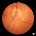 |
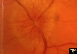
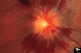
A201 Disc Swelling with Big Blind Spot Syndrome | Blind spot larger than could be explained by visible edema. Subretinal white dots probably indicate margin of blind spot. Anatomy: Optic disc; Retina. Pathology: Unknown. Disease/ Diagnosis: Big blind spot syndrome. Clinical: symptoms: photosias, blurred vision signs: Disc swelling; white spots in t... | Image |
| 2 |
 |
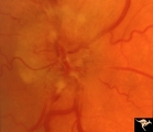
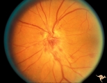
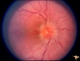
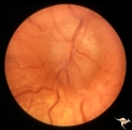
A303 Disc Swelling, Chorioretinal Disease | Neovascular net. Disc swelling with peripapillary neo-vascularization with subretinal hemorrhage. Anatomy: Optic disc; Retina. | Image |
| 3 |
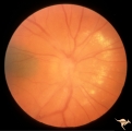 |
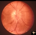
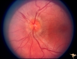
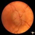
A404 Disc Swelling, Vitreous Effects | Disc elevation (swelling) and vitritis. Posterior vitreous detachment with vitritis. Incidental choroidal nevus. Anatomy: Optic disc; Vitreous; Retina. Pathology: Posterior vitreal detachment, disc swelling, and vitritis-horoidal nevus. Disease/ Diagnosis: Vitreal detachment from optic disc; choroid... | Image |
| 4 |
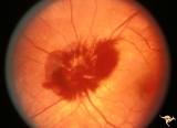 |
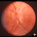
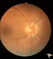
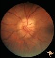
A407 Disc Swelling, Vitreous Effects | Prepapillary hemorrhage. Partial posterior vitreous detachment in myopic patient. Reference: Katz B, Hoyt WF. Intrapapillary and peripapillary hemorrhage in young patients with incomplete posterior vitreous detachment. Signs of vitreopapillary traction. Ophthalmology. 1995 Feb;102(2):349-54. Anatomy... | Image |
| 5 |
 |
B102 Disc Swelling, Ischemic Papillopathies, AION | Ischemic swelling. March 2, 1978. Same patient as B1_03. Anatomy: Optic disc. Pathology: Axoplasmic stasis due to ischemia. Disease/ Diagnosis: AION. Clinical: Diabetic with optic disc swelling and visual loss. | Image |
| 6 |
 |
B103 Disc Swelling, Ischemic Papillopathies, AION | Ischemic swelling. 50 year old woman, 12 days after a viral illness. Nasal nerve fiber layer bundle visual field defect. Anatomy: Optic disc. Pathology: Axoplasmic stasis due to ischemia. Disease/ Diagnosis: AION. Clinical: Visual loss after viral illness. | Image |
| 7 |
 |
B104 Disc Swelling, Ischemic Papillopathies, AION | Ischemic swelling. 57 year old man. Anatomy: Optic disc. Pathology: Axoplasmic stasis due to ischemia. Disease/ Diagnosis: AION. Clinical: Visual loss. | Image |
| 8 |
 |
B108 Disc Swelling, Ischemic Papillopathies, AION | Pallid ischemic swelling. Woman with vasculitis. Anatomy: Optic disc. Pathology: Axoplasmic stasis due to ischemia. Disease/ Diagnosis: AION. Clinical: Visual loss. | Image |
| 9 |
 |
B109 Disc Swelling, Ischemic Papillopathies, AION | Ischemic swelling. Patient was diabetic. April 18, 1978. Same patient as B1-2. Anatomy: Optic disc. Pathology: Axoplasmic stasis due to ischemia. Disease/ Diagnosis: AION. Clinical: Diabetic with disc swelling and visual loss. | Image |
| 10 |
 |
B205 Disc Swelling, Diabetic Papillopathy | Bilateral diabetic papillopathy. Girl. Left eye. Pair with B2_06. Anatomy: Optic disc. Pathology: Axoplasmic stasis due to ischemia. Disease/ Diagnosis: Diabetic papillopathy. Clinical: Visual loss with recovery. | Image |
| 11 |
 |
B206 Disc Swelling, Diabetic Papillopathy | Bilateral diabetic papillopathy. Girl. Right eye. Pair with B2_05. Anatomy: Optic disc. Pathology: Axoplasmic stasis due to ischemia. Disease/ Diagnosis: Diabetic papillopathy. Clinical: Visual loss with recovery. | Image |
| 12 |
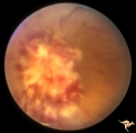 |
B405 Disc Swelling, Radiation Papillopathy | Bilateral blindness 6 months post radiation for malignant glioma of left hemisphere. Left eye. Marked white exudation probably represents necrosis of swollen disc tissue. Japanese patient. Anatomy: Optic disc. Pathology: Axoplasmic stasis due to ischemia. Disease/ Diagnosis:Radiation papillopathy; O... | Image |
| 13 |
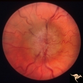 |
Bilateral Chronic Papilledema | Left eye. Frisen's stage 5. Patient with long standing aqueductal stenosis. Bilateral Chronic Papilledema. Man. Anatomy: Optic disc. Pathology: Papilledema. Disease/Diagnosis: Papilledema from aqueductal stenosis. | Image |
| 14 |
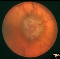 |
Bilateral Chronic Papilledema | Right eye. Frisen's stage 5. Patient with long standing aqueductal stenosis. Bilateral Chronic Papilledema. Man. Anatomy: Optic disc. Pathology: Papilledema. Disease/Diagnosis: Papilledema from aqueductal stenosis. | Image |
| 15 |
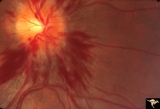 |
Bilateral Hemorrhagic Papilledema from Saggital Sinus Thrombosis | Left eye. 20 year old woman on oral contraceptives. Bilateral hemorrhagic Papilledema from sagittal sinus thrombosis. Anatomy: Optic disc. Pathology: Papilledema; hemorrhagic papilledema. Disease/Diagnosis: Superior saggital sinus thrombosis due to BCP use. Clinical notes: Chronic headache. | Image |
| 16 |
 |
Bilateral Hemorrhagic Papilledema from Saggital Sinus Thrombosis | Right eye. 20 year old woman on oral contraceptives. Bilateral hemorrhagic Papilledema from sagittal sinus thrombosis. Anatomy: Optic disc. Pathology: Papilledema; hemorrhagic papilledema. Disease/Diagnosis: Superior sagittal sinus thrombosis due to BCP use. Clinical notes: Chronic headache. | Image |
| 17 |
 |
Bilateral Papilledema | Right eye. Bilateral Papilledema in patient with cardiopulmonary insufficiency. Woman. Anatomy: Optic disc. Pathology: Papilledema. Disease/Diagnosis: cardiopulmonary insufficiency causing intracranial hypertension. Clinical notes: headache. | Image |
| 18 |
 |
Bilateral Papilledema | Left eye. Bilateral Papilledema in patient with cardiopulmonary insufficiency. Woman. Anatomy: Optic disc. Pathology: Papilledema. Disease/Diagnosis: Cardiopulmonary insufficiency causing intracranial hypertension. Clinical notes: Headache. | Image |
| 19 |
 |
Bilateral Papilledema | Left eye. Chronic Bilateral Papilledema. Anatomy: Optic disc. Pathology: Chronic bilateral papilledema. Disease/Diagnosis: Pseudotumor long standing. Clinical notes: Chronic headache; Obesity. | Image |
| 20 |
 |
Bilateral Papilledema | Left eye. Bilateral Papilledema from vitamin A toxicity in young girl. Anatomy: Optic disc. Pathology: Bilateral papilledema. Disease/Diagnosis: Pseudotumor due to vitamin A toxicity in a young girl. Clinical notes: Headache. | Image |
| 21 |
 |
Bilateral Papilledema | Right eye. Bilateral Papilledema from vitamin A toxicity in young girl. Anatomy: Optic disc. Pathology: Bilateral papilledema. Disease/Diagnosis: Pseudotumor due to vitamin A toxicity in a young girl. Clinical notes: Headache. | Image |
| 22 |
 |
Bilateral Papilledema | Chronic Bilateral Papilledema. Anatomy: Optic disc. Pathology: Chronic bilateral papilledema. Disease/Diagnosis: Pseudotumor long standing. Clinical notes: Chronic headache; Obesity. | Image |
| 23 |
 |
Bilateral Papilledema from Pseudotumor | Right eye. Pseudotumor syndrome. Multiple endocrine adenomas. Woman. Anatomy: Optic disc. Pathology: Bilateral papilledema. Disease/Diagnosis: Pseudotumor associated with multiple endocrine adenomas. Clinical notes: Headache; Obesity. | Image |
| 24 |
 |
Bilateral Papilledema in Pseudotumor | Left eye. Pseudotumor syndrome. Multiple endocrine adenomas. Woman. Anatomy: Optic disc. Pathology: Bilateral papilledema. Disease/Diagnosis: Pseudotumor associated with multiple endocrine adenomas. Clinical notes: Headache; Obesity. | Image |
| 25 |
 |
Bilateral Papilledema with Cyanotic Heart Disease | Bilateral Papilledema with cyanotic heart disease in a young boy. Anatomy: Optic disc. Pathology: Papilledema. Disease/Diagnosis: Pseudotumor due to cyanotic heart disease. Clinical notes: Young boy with clubbing. | Image |