Best known for his world-renowned neuro-ophthalmology unit based at the University of California, San Francisco, William Hoyt, MD collected here more than 850 of his best images covering a wide range of disorders.
William F. Hoyt, MD, Professor Emeritus of Ophthalmology, Neurology and Neurosurgery, Department of Ophthalmology, University of California, San Francisco.
NOVEL: https://novel.utah.edu/
TO
Filters: Collection: "ehsl_novel_wfh"
| Title | Description | Type | ||
|---|---|---|---|---|
| 1 |
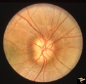 |
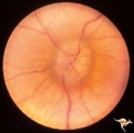
Buried Drusen | 7 year old boy with pseudo papilledema from buried drusen. Note the lumpy contour of the disc margin. Also note the surrounding ring-like light reflex that is optically perfect and indicates absence of edema spreading onto the surrounding retina. Anatomy: Optic disc. Pathology: Drusen of the optic d... | Image |
| 2 |
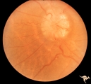 |
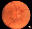
Chronic Papilledema with Pseudo Drusen | Right eye. Meningioma. Pseudo drusen from chronic papilledema. Woman. Anatomy: Optic disc Pathology: Papilledema Disease/Diagnosis: Chronic papilledema with pseudo drusen | Image |
| 3 |
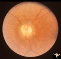 |
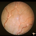
Chronic Papilledema with Pseudo Drusen | Left eye. Meningioma. Pseudo drusen from chronic papilledema. The patient's meningioma had blinded her left eye and caused chronic elevated intracranial pressure. Woman. Anatomy: Optic disc Pathology: Papilledema Disease/Diagnosis: Chronic papilledema with pseudo drusen | Image |
| 4 |
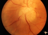 |
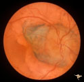
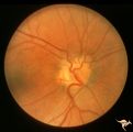
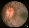
F107 Metastatic Breast Cancer to the Disc | Metastatic breast cancer to the disc. Notice mass on inferior portion of disc. Also notice tangled capillary dilation within the mass indicating infiltration. This disc tumor was radiated. It disappeared leaving a pale flat atrophic nerve. The patient died. Histologic study of the eye revealed metas... | Image |
| 5 |
 |
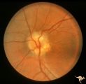
Buried Drusen with Choroidal Retinal Scar | Right eye: Buried drusen; probable complication of peripapillary hemorrhage at 7:00. Anatomy: Optic disc. Pathology: Drusen of the optic disc. Disease/Diagnosis: Drusen of the optic disc. Clinical notes: Enlarged blind spot. | Image |
| 6 |
 |
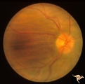
Drusen with Sub-retinal Neovascular Net | Buried drusen with sub-retinal neovascular net. There may be retinoschisis as well. Anatomy: Optic disc. Pathology: Drusen plus neovascularization at the border of the optic disc. Disease/Diagnosis: Drusen of the optic disc. Clinical: Patient has very large blind spot and impaired central vision. | Image |
| 7 |
 |
H29 Dysplasia with Hypoplasia (Elevated Dysplasia with Anomalous Vessels) | Right eye. Dysplasia with anomalous cilioretinal arterioles. Central retinal artery may be absent. Same patient as H_30. Anatomy: Optic disc. Pathology: Dysplasia of the optic disc. Disease/ Diagnosis: Elevated dysplasia with hypoplasia. | Image |
| 8 |
 |
H30 Dysplasia with Hypoplasia (Elevated Dysplasia with Anomalous Vessels) | Left eye. Elevated dysplasia with anomalous cilioretinal vessels. Central retinal artery may be absent. Same patient as H_29. Anatomy: Optic disc. Pathology: Dysplasia of the optic disc. Disease/ Diagnosis: Elevated dysplasia with hypoplasia. | Image |
| 9 |
 |
Post Papilledema with Choroidal Folds | Right eye. Post papilledema with choroidal folds due to brain tumor. Anatomy: Optic disc. Pathology: Post papilledema. Disease/Diagnosis: Post papilledema with choroidal folds. | Image |
| 10 |
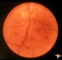 |
Slow Flow (Chronic Hypoxic) Retinopathy | Slow flow (chronic hypoxic) retinopathy from macroglobulanemia. Note the dot and blot hemorrhages. Anatomy: Retina. Pathology: Macroglobulanemia. Disease/Diagnosis: Slow flow (chronic hypoxic) retinopathy from macroglobulanemia. | Image |
| 11 |
 |
Unilateral Papilledema | Right eye. Atrophic nerve right eye. Large falx meningioma. True Foster Kennedy Syndrome. Anatomy: Optic disc. Pathology: Chronic papilledema; optic atrophy. Disease/Diagnosis: Meningioma causing Foster-Kennedy Syndrome. Clinical: Visual loss one eye; Transient visual obscuration other eye. | Image |
| 12 |
 |
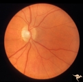
Unilateral Papilledema | Left eye. Left eye has papilledema. Large falx meningioma. True Foster Kennedy Syndrome. Anatomy: Optic disc. Pathology: Chronic papilledema; optic atrophy. Disease/Diagnosis: Meningioma causing Foster-Kennedy Syndrome. Clinical: Visual loss one eye; transient visual obscuration other eye. | Image |
| 13 |
 |
Unilateral Papilledema | Right eye. Has slight disc blur. Asymmetric papilledema. 35 year old woman. Anatomy: Optic disc. Pathology: Unilateral papilledema. Disease/Diagnosis: Idiopathic intracranial hypertension (pseudotumor cerebri). Clinical: Gaze evoked blindness. | Image |
| 14 |
 |
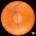
Unilateral Papilledema | Left eye. This eye has papilledema. 35 year old woman. Anatomy: Optic disc. Pathology: Unilateral papilledema. Disease/Diagnosis: Idiopathic intracranial hypertension (pseudotumor cerebri). Clinical: Gaze evoked blindness. | Image |
| 15 |
 |
Vascular Disc Anomalies - Prepapillary Arterial Convolutions | Prepapillary arterial convolutions. Left eye. Man. Anatomy: Optic disc. Pathology: Congenital prepapillary arterial convolutions. Disease/Diagnosis: Congenital arterial vascular anomaly. Clinical: Asymptomatic. | Image |
| 16 |
 |
Buried Drusen | 5 year old boy. Bilateral buried drusen. Notice the lumpy nasal disc elevation. This patient had a twin brother whose optic disc drusen were exposed. Anatomy: Optic disc. Pathology: Drusen of the optic disc. Disease/Diagnosis: Drusen of the optic disc. Clinical notes: Normally functioning eye with ... | Image |
| 17 |
 |
Buried Drusen | 5 year old boy. Bilateral buried drusen. Notice the lumpy nasal disc elevation. This patient had a twin brother whose optic disc drusen were exposed. Anatomy: Optic disc. Pathology: Drusen of the optic disc. Disease/Diagnosis: Drusen of the optic disc. Clinical notes: Normally functioning eye with ... | Image |
| 18 |
 |
Chronic Papilledema with Pseudo Drusen | Left eye. Chronic papilledema with pseudo drusen due to cerebral pontine angle tumor. Anatomy: Optic disc. Pathology: Papilledema Disease/Diagnosis: Chronic papilledema with pseudo drusen. | Image |
| 19 |
 |
Chronic Papilledema with Pseudo Drusen | Right eye. Chronic papilledema with pseudo drusen due to cerebral pontine angle tumor. Anatomy: Optic disc Pathology: Papilledema Disease/Diagnosis: Chronic papilledema with pseudo drusen | Image |
| 20 |
 |
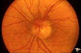
E09 Disc Swelling with Central Vein Occlusion | Chronic disc swelling due to CRVO. Anatomy: Optic disc; Retina. Pathology: Central retinal vein occlusion. Disease/ Diagnosis: Disc swelling due to central retinal vein occlusion. | Image |
| 21 |
 |
H25 Dysplasia with Hypoplasia (Elevated Dysplasia with Anomalous Vessels) | Right eye. Elevated dysplasia with anomalous blood vessel pattern and peri-papillary choroidal malformation. Same patient as H_26. Anatomy: Optic disc. Pathology: Dysplasia of the optic disc. Disease/ Diagnosis: Elevated dysplasia with hypoplasia. | Image |
| 22 |
 |
H26 Dysplasia with Hypoplasia (Elevated Dysplasia with Anomalous Vessels) | Left eye. Dysplasia with grossly anomalous vascular pattern. Elevated dysplasia. Same patient as H_25. Anatomy: Optic disc. Pathology: Dysplasia of the optic disc. Disease/ Diagnosis: Elevated dysplasia with hypoplasia. | Image |
| 23 |
 |
C302 Nodular Papillopathies (Sarcoid) | Perivenous Inflammatory Cuffing in a Patient with Proven Sarcoid. Left eye. Pair with C3_01. Anatomy: Retina. Pathology: Axoplasmic stasis due to sarcoid infiltration with retinal venous exudation? Disease/ Diagnosis: Sarcoid papillopathy with perivenous inflammatory disease. Clinical: Unknown? | Image |
| 24 |
 |
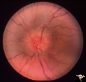
C301 Nodular Papillopathies (Sarcoid) | Disc swelling. Sarcoid papillopathy. Note infiltrative nodule at 9:00 on the disc.The patient had proven sarcoid. Perivenous inflammatory cuffing visible on image C3_02. Right eye. Pair with C3_02. Anatomy: Optic disc; Retina. Pathology: Axoplasmic stasis due to sarcoid infiltration. Disease/ Diagn... | Image |
| 25 |
 |
Bilateral Papilledema | Left eye. Bilateral Papilledema with hypoparathyroidism. Woman. Anatomy: Optic disc. Pathology: Papilledema. Papilledema with hypopararthyroidism. | Image |