Best known for his world-renowned neuro-ophthalmology unit based at the University of California, San Francisco, William Hoyt, MD collected here more than 850 of his best images covering a wide range of disorders.
William F. Hoyt, MD, Professor Emeritus of Ophthalmology, Neurology and Neurosurgery, Department of Ophthalmology, University of California, San Francisco.
NOVEL: https://novel.utah.edu/
TO
Filters: Collection: "ehsl_novel_wfh"
| Title | Description | Type | ||
|---|---|---|---|---|
| 1 |
 |
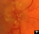
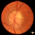
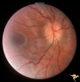
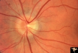
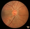
B101 Disc Swelling, Ischemic Papillopathies, AION | Pallid swelling in course of acute AION. 48 year old man who developed disc swelling after a flu like illness, then developed AION. Anatomy: Optic disc. Pathology: Axoplasmic stasis due to ischemia. Disease/ Diagnosis: AION. Clinical: Visual loss after flu-like illness. | Image |
| 2 |
 |
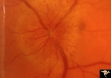
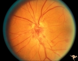
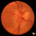
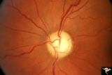
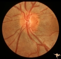
B102 Disc Swelling, Ischemic Papillopathies, AION | Ischemic swelling. March 2, 1978. Same patient as B1_03. Anatomy: Optic disc. Pathology: Axoplasmic stasis due to ischemia. Disease/ Diagnosis: AION. Clinical: Diabetic with optic disc swelling and visual loss. | Image |
| 3 |
 |
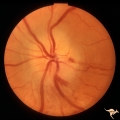
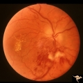
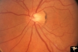
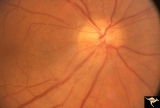
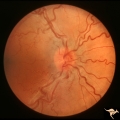
B103 Disc Swelling, Ischemic Papillopathies, AION | Ischemic swelling. 50 year old woman, 12 days after a viral illness. Nasal nerve fiber layer bundle visual field defect. Anatomy: Optic disc. Pathology: Axoplasmic stasis due to ischemia. Disease/ Diagnosis: AION. Clinical: Visual loss after viral illness. | Image |
| 4 |
 |
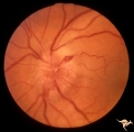
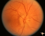
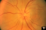
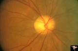
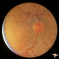
B104 Disc Swelling, Ischemic Papillopathies, AION | Ischemic swelling. 57 year old man. Anatomy: Optic disc. Pathology: Axoplasmic stasis due to ischemia. Disease/ Diagnosis: AION. Clinical: Visual loss. | Image |
| 5 |
 |
B105 Disc Swelling, Ischemic Papillopathies, AION | Pallid ischemic swelling. 48 year old woman, flight attendant. Anatomy: Optic disc. Pathology: Axoplasmic stasis due to ischemia. Disease/ Diagnosis: AION. Clinical: Visual loss. | Image |
| 6 |
 |
B106 Disc Swelling, Ischemic Papillopathies, AION | Red ischemic swelling. 49 year old man. Anatomy: Optic disc. Pathology: Axoplasmic stasis due to ischemia. Disease/ Diagnosis: AION. Clinical: Visual loss. | Image |
| 7 |
 |
B107 Disc Swelling, Ischemic Papillopathies, AION | Pallid ischemic swelling. 41 year old man. Anatomy: Optic disc. Pathology: Axoplasmic stasis due to ischemia. Disease/ Diagnosis: AION. Clinical: Viusal loss. | Image |
| 8 |
 |
B108 Disc Swelling, Ischemic Papillopathies, AION | Pallid ischemic swelling. Woman with vasculitis. Anatomy: Optic disc. Pathology: Axoplasmic stasis due to ischemia. Disease/ Diagnosis: AION. Clinical: Visual loss. | Image |
| 9 |
 |
B109 Disc Swelling, Ischemic Papillopathies, AION | Ischemic swelling. Patient was diabetic. April 18, 1978. Same patient as B1-2. Anatomy: Optic disc. Pathology: Axoplasmic stasis due to ischemia. Disease/ Diagnosis: AION. Clinical: Diabetic with disc swelling and visual loss. | Image |
| 10 |
 |
B110 Disc Swelling, Ischemic Papillopathies, AION | Pallid ischemic swelling with intraretinal exudates near the macula and a ""cotton wool"" infarct below the disc. 38 year old man. Diabetic. 20/60 vision. Altitudinal visual field defect. Anatomy: Optic disc. Pathology: Axoplasmic stasis due to ischemia. Disease/ Diagnosis: AION. Clinical: Diabetic ... | Image |
| 11 |
 |
B111 Disc Swelling, Ischemic Papillopathies, AION | Acute AION. Anatomy: Optic disc. Pathology: Axoplasmic stasis due to ischemia. Disease/ Diagnosis: AION. Clinical: Visual loss. | Image |
| 12 |
 |
B112 Disc Swelling, Ischemic Papillopathies, AION | Arterioles are narrowing in resolution phase from AION. Patient had a superior altitudinal visual field defect. 20 year old man. Anatomy: Optic disc. Pathology: Axoplasmic stasis due to ischemia. Disease/ Diagnosis: AION. Clinical: Visual loss. | Image |
| 13 |
 |
B113 Disc Swelling, Ischemic Papillopathies, AION | 57 year old woman with AION. Anatomy: Optic disc. Pathology: Axoplasmic stasis due to ischemia. Disease/ Diagnosis: AION. Clinical: Visual loss. | Image |
| 14 |
 |
B114 Disc Swelling, Ischemic Papillopathies, AION | AION in a disc with an optic cup. Extraordinary exception with AION. Note ischemic vascular changes in disc surface. Anatomy: Optic disc. Pathology: Axoplasmic stasis due to ischemia. Disease/ Diagnosis: AION. Clinical: Visual loss. | Image |
| 15 |
 |
B115 Disc Swelling, Ischemic Papillopathies, AION | Normal eye in patient who later developed AION. Note generous optic cup. June 2, 1991. Same patient as B1_16b. Anatomy: Optic disc. Pathology: Normal. Clinical: Asymptomatic. | Image |
| 16 |
 |
B116 Disc Swelling, Ischemic Papillopathies, AION | Typical AION in disc with optic cup. December 23, 2996. 5 years later in same patient as B1_15a. Anatomy: Optic disc. Pathology: Axoplasmic stasis due to ischemia. Disease/ Diagnosis: AION. Clinical: Visual loss. | Image |
| 17 |
 |
C115 Papillitis, Retrobulbar Neuritis | Demyelinative optic neuropathy with mild disc swelling. This eye had a large central scotoma. Note the bland disc margin swelling from 2:00 to 4:00. This swelling constitutes spill over edema from the main focus of the neuritis which lies behind the eyeball. Visual acuity was 2200. Anatomy: Optic di... | Image |
| 18 |
 |
End Stage Leber Optic Neuropathy | End stage Leber's Optic Neuropathy. Severe diffuse pallor. Left eye. Pair with 15a. Anatomy: Optic disc. Pathology: Optic neuropathy. Disease/ Diagnosis: Leber's optic neuropathy. Clinical: Blindness. | Image |
| 19 |
 |
IB114a Post Ischemic (AION) Cupless Atrophy | 1991, acute AION in a disc with a cup, pair with IB1_14b. Anatomy: Optic disc. Pathology: Post ischemic (AION) cupless atrophy. Disease/ Diagnosis: Post ischemic (AION) cupless atrophy. Clinical: Visual loss. | Image |
| 20 |
 |
IB114b Post Ischemic (AION) Atrophy in a Disc with a Cup | 1996, same as IB1_14a five years later reveals pallor, arteriole narrowing and optic cup. Anatomy: Optic disc. Pathology: Post ischemic (AION) cupless atrophy. Disease/ Diagnosis: Post ischemic (AION) cupless atrophy. Clinical: Visual loss. | Image |
| 21 |
 |
IE01 Acute Leber Optic Neuropathy | Pseudo edema with peripapillary microangiopathy in a brother of boy with Leber Optic Neuropathy. Not pale nerve yet. Right eye. Same patient as IE_02a and IE_02b. August 7, 1979. Anatomy: Optic disc. Pathology: Optic neuropathy. Disease/ Diagnosis: Optic neuropathy. Clinical: Asymptomatic | Image |
| 22 |
 |
IE02a Acute Leber Optic Neuropathy | Acute Leber Optic Neuropathy, Left eye. Same patient as IE_01 and IE_02b. August 7, 1979. Anatomy: Optic disc. Pathology: Optic neuropathy. Disease/ Diagnosis: Leber's optic neuropathy. Clinical: Asymptomatic. | Image |
| 23 |
 |
IE02b Acute Leber Optic Neuropathy | Acute Leber Optic Neuropathy. Formation of hemorrhage one year after IE_02a. At this time, he was beginning to lose central vision. .Note thinning of the nerve fiber layer temporally, 3:00 - 4:00 Pair with IE_02a. March 26, 1980. Anatomy: Optic disc. Pathology: Optic neuropathy. Disease/ Diagnosis: ... | Image |
| 24 |
 |
IE03 Acute Leber Optic Neuropathy | Acute stage of Leber optic neuropathy with microangiopathy and peripapillary nerve fiber layer thickening. The temporal nerve fiber layer is already showing atrophy. Central vision is grossly reduced. 1971. Anatomy: Optic disc. Pathology: Optic neuropathy. Disease/ Diagnosis: Leber's optic neuropath... | Image |
| 25 |
 |
IE04 Acute Leber Optic Neuropathy | Microangiopathy without visual loss in a patient with acute Leber's optic neuropathy in the left eye. Pair with IE_05. Anatomy: Optic disc. Pathology: Optic neuropathy. Disease/ Diagnosis: Leber's optic neuropathy. Clinical: Central vision loss. | Image |