Best known for his world-renowned neuro-ophthalmology unit based at the University of California, San Francisco, William Hoyt, MD collected here more than 850 of his best images covering a wide range of disorders.
William F. Hoyt, MD, Professor Emeritus of Ophthalmology, Neurology and Neurosurgery, Department of Ophthalmology, University of California, San Francisco.
NOVEL: https://novel.utah.edu/
TO
Filters: Collection: "ehsl_novel_wfh"
| Title | Description | Type | ||
|---|---|---|---|---|
| 1 |
 |
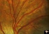
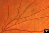
Multiple Sclerosis Slits and Thinning in Peripapillary (Retinal) Nerve Riber Layer | Left eye. Upper arcuate nerve fiber layer contains multiple low density slits. These indicate nerve fiber loss. Anatomy: Peripapillary nerve fiber layer. Pathology: Slit-like atrophy. Disease/Diagnosis: Multiple sclerosis optic neuropathy. Clinical: No symptoms. | Image |
| 2 |
 |
Multiple Sclerosis Slits and Thinning in Peripapillary (Retinal) Nerve Riber Layer | Multiple slit defect in the superior arcuate nerve fiber layer. Anatomy: Peripapillary nerve fiber layer. Pathology: Slit-like atrophy. Disease/Diagnosis: Multiple sclerosis optic neuropathy. Clinical: No symptoms. | Image |
| 3 |
 |
Multiple Sclerosis Slits and Thinning in Peripapillary (Retinal) Nerve Riber Layer | Multiple slit defect in the superior arcuate nerve fiber layer. Anatomy: Peripapillary nerve fiber layer. Pathology: Slit-like atrophy. Disease/Diagnosis: Multiple sclerosis optic neuropathy. Clinical: No symptoms. | Image |
| 4 |
 |
Multiple Sclerosis Slits and Thinning in Peripapillary (Retinal) Nerve Riber Layer | Multiple slit defect in the superior arcuate nerve fiber layer. Pair with IIB2_6b. Anatomy: Peripapillary nerve fiber layer. Pathology: Slit-like atrophy. Disease/Diagnosis: Multiple sclerosis optic neuropathy. Clinical: No symptoms. | Image |
| 5 |
 |
Multiple Sclerosis Slits and Thinning in Peripapillary (Retinal) Nerve Riber Layer | Multiple slit like defects in the inferior arcuate nerve fibers. Pair with IIB2_3b. Anatomy: Peripapillary nerve fiber layer. Pathology: Slit-like atrophy. Disease/Diagnosis: Multiple sclerosis optic neuropathy. Clinical: No symptoms. | Image |
| 6 |
 |
Multiple Sclerosis Slits and Thinning in Peripapillary (Retinal) Nerve Riber Layer | Multiple slit and wedge defects in the nerve fiber layer. Pair with IIB2_3a. Anatomy: Peripapillary nerve fiber layer. Pathology: Slit-like atrophy. Disease/Diagnosis: Multiple sclerosis optic neuropathy. Clinical: No symptoms. | Image |
| 7 |
 |
Multiple Sclerosis Slits and Thinning in Peripapillary (Retinal) Nerve Riber Layer | Multiple slit defect in the superior arcuate nerve fiber layer. Magnified. Pair with IIB2_6a. Anatomy: Peripapillary nerve fiber layer. Pathology: Slit-like atrophy. Disease/Diagnosis: Sclerosis optic neuropathy. Clinical: No symptoms. | Image |
| 8 |
 |
Multiple Sclerosis Slits and Thinning in Peripapillary (Retinal) Nerve Riber Layer | Multiple slit defect in the superior arcuate nerve fiber layer in a 13 year old boy. Right eye. Pair with IIB2_7a. Anatomy: Peripapillary nerve fiber layer. Pathology: Slit-like atrophy. Disease/Diagnosis: Multiple sclerosis optic neuropathy. Clinical: No symptoms. | Image |
| 9 |
 |
Multiple Sclerosis Slits and Thinning in Peripapillary (Retinal) Nerve Riber Layer | Multiple slit defect in the superior arcuate nerve fiber layer in a 13 year old boy. Pair with IIB2_7b. Anatomy: Peripapillary nerve fiber layer. Pathology: Slit-like atrophy. Disease/Diagnosis: Multiple sclerosis optic neuropathy. Clinical: No symptoms. | Image |
| 10 |
 |
Multiple Sclerosis Slits and Thinning in Peripapillary (Retinal) Nerve Riber Layer | Need magnification - Left eye - Inferior arcuate nerve fiber slits. Pairs with IIB2_01b & IIB2_01c. Anatomy: Peripapillary nerve fiber layer. Pathology: Slit-like atrophy. Disease/Diagnosis: Multiple sclerosis optic neuropathy. Clinical: No symptoms. | Image |
| 11 |
 |
Multiple Sclerosis Slits and Thinning in Peripapillary (Retinal) Nerve Riber Layer | Need magnification - Left eye - Inferior arcuate nerve fiber slits. Pairs with IIB2_01a & IIB2_01c. Anatomy: Peripapillary nerve fiber layer. Pathology: Slit-like atrophy. Disease/Diagnosis: Multiple sclerosis optic neuropathy. Clinical: No symptoms. | Image |
| 12 |
 |
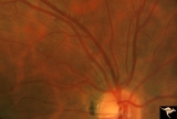
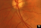
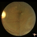
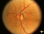
F202 Optic Nerve Sheath Meningioma | Optic nerve sheath meningioma. Note optociliary vein at 3:00. The disc is atrophic. Anatomy: Optic disc. Pathology: Chronic optic disc swelling caused by optic nerve sheath meningioma. Disease/ Diagnosis: Chronic optic disc swelling caused by optic nerve sheath meningioma. | Image |
| 13 |
 |
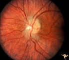
F203 Optic Nerve Sheath Meningioma | Optic nerve sheath meningioma. Note optociliary vessels on the disc. The disc is partially atrophic and blurred by previous edema. The cause of the choroidal scar was not determined. Anatomy: Optic disc. Pathology: Chronic optic disc swelling caused by optic nerve sheath meningioma. DIsease/ Diagnos... | Image |
| 14 |
 |
F204 Optic Nerve Sheath Meningioma | Optic disc swelling due to meningioma. Notice choroidal folds through the macula of left eye. Anatomy: Optic disc. Pathology: Chronic optic disc swelling caused by optic nerve sheath meningioma. Disease/ Diagnosis: Chronic optic disc swelling caused by optic nerve sheath meningioma. | Image |
| 15 |
 |
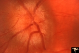
Diffuse Atrophy | Nerve fiber appearance about 6 weeks after indirect injury to optic nerve. Note near total absence of nerve fiber reflexes. Photo shows remaining streaks of inferior arcuate nerve fiber membranes dissolving into nothing. 1972. Anatomy: Optic disc. Pathology: Optic nerve injury. Disease/Diagnosis: Op... | Image |
| 16 |
 |
Multiple Sclerosis Slits and Thinning in Peripapillary (Retinal) Nerve Riber Layer | Need magnification - Left eye - Peculiar punctate dotted surface of internal limiting membrane reflexes. Pairs with IIB2_01a & IIB2_02b. Anatomy: Peripapillary nerve fiber layer. Pathology: Slit-like atrophy. Disease/Diagnosis: Multiple sclerosis optic neuropathy. Clinical: No symptoms. | Image |
| 17 |
 |
Diffuse Atrophy - Evolution of Optic Disc Palor After Optic Nerve Transection | Injury on December 8, 1978. Evolution of optic disc pallor after optic nerve transection. Woman having rhinoplasty suffered optic nerve transection. Left eye. Photo taken January 11, 1979 - 33 days post accident. Note superior and inferior arcuate nerve fiber bundles are thinned. Optic disc shows s... | Image |
| 18 |
 |
Diffuse Atrophy - Evolution of Optic Disc Palor After Optic Nerve Transection | Injury on December 8, 1978. Evolution of optic disc pallor after optic nerve transection. Woman having rhinoplasty suffered optic nerve transection. Left eye. Photo taken January 18, 1979 - 40 days post accident. Retinal nerve fiber layer appears thinner and disc is paler. Anatomy: Optic disc. Patho... | Image |
| 19 |
 |
Diffuse Atrophy - Evolution of Optic Disc Palor After Optic Nerve Transection | Injury on December 8, 1978. Evolution of optic disc pallor after optic nerve transection. Woman having rhinoplasty suffered optic nerve transection. One day after nerve transection. Note dilated veins. Left eye. Photo taken December 9, 1978. Anatomy: Optic disc. Pathology: Total retrograde optic atr... | Image |
| 20 |
 |
Diffuse Atrophy - Evolution of Optic Disc Palor After Optic Nerve Transection | Injury on December 8, 1978. Evolution of optic disc pallor after optic nerve transection. Woman having rhinoplasty suffered optic nerve transection. Left eye. Photo taken February 14, 1979 - 65 days post accident. Optic disc is completely pale. All evidence of retinal nerve fiber layer is gone. Anat... | Image |
| 21 |
 |
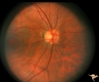
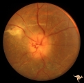
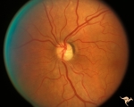
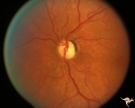
F201 Optic Nerve Sheath Meningioma | Right eye. Woman with ophthalmoplegia proptosis for 14 years. Visual field reduced due to optic nerve sheath meningioma. Notice large optociliary vessel temporally. Anatomy: Optic disc. Pathology: Chronic optic disc swelling caused by optic nerve sheath meningioma. Disease/ Diagnosis: Chronic optic ... | Image |
| 22 |
 |
Diffuse Atrophy - Evolution of Optic Disc Palor After Optic Nerve Transection | Evolution of optic disc pallor after optic nerve transection. Normal Right eye. Photo taken December 9, 1978. Anatomy: Optic disc. Pathology: Total retrograde optic atrophy. Disease/Diagnosis: Transection of the optic nerve. Clinical: Blindness. | Image |
| 23 |
 |
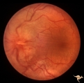
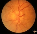
F2b01 Optic Nerve Glioma | Left eye. Woman with optic nerve glioma. Anatomy: Optic disc. Pathology: Optic nerve swelling secondary to retrobulbar optic glioma. Disease/ Diagnosis: Optic nerve glioma. | Image |
| 24 |
 |
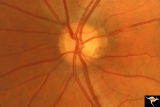
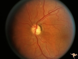
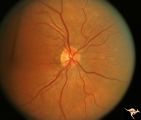
Segmental Atrophy - Hemianopic (Band) Atrophy from Eight Optic Tract Injury | Segmental Atrophy - Band atrophy from right optic tract injury. This eye has a nasal hemianopia. Its disc shows temporal pallor with an intact nasal nerve fiber layer. Pair with IIA2C_7b. Anatomy: Optic disc. Pathology: Right optic tract injury. Disease/Diagnosis: Homonymous hemioptic atrophy. Clini... | Image |
| 25 |
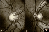 |
Segmental Atrophy - Hemianopic (Band) Atrophy | Segmental Atrophy - Band atrophy. Shows band atrophy in left disc with preserved upper and lower arcuate nerve fiber bundles. Right disc has thinning of both upper and lower arcuate nerve fiber bundles, temporal pallor, and an intact nasal nerve fiber layer. 1972. Anatomy: Optic disc. Pathology: Rig... | Image |