Best known for his world-renowned neuro-ophthalmology unit based at the University of California, San Francisco, William Hoyt, MD collected here more than 850 of his best images covering a wide range of disorders.
William F. Hoyt, MD, Professor Emeritus of Ophthalmology, Neurology and Neurosurgery, Department of Ophthalmology, University of California, San Francisco.
NOVEL: https://novel.utah.edu/
TO
Filters: Collection: "ehsl_novel_wfh"
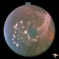
1 - 25 of 11
| Title | Description | Type | ||
|---|---|---|---|---|
| 1 |
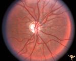 |
Neurofibromatosis-1 | Optic atrophy and hypoplasia of the optic disc associated with chiasmal glioma in a patient with NF-1. Anatomy: Optic disc. Pathology: Chiasmal glioma; Optic atrophy; Hypoplasia. Disease/Diagnosis: Neurofibromatosis type 1. Clinical: Proptosis; Blindness. | Image |
| 2 |
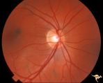 |
Neurofibromatosis-1 | Normal appearing optic disc with dark pigmented choroidal nevi. The patient had NF-1 and had a subclinical optic glioma on the left eye. This is the right eye. Anatomy: Optic disc. Pathology: Choroidal nevus. Disease/Diagnosis: Neurofibromatosis type 1. Clinical: No visual symptoms. | Image |
| 3 |
 |
Neurofibromatosis-1 | Retinal microvascular malformations in NF-1 located between the disc and the macula. Anatomy: Retina. Pathology: Retinal microvascular malformations. Disease/Diagnosis: Neurofibromatosis type 1. Clinical: No visual symptoms. | Image |
| 4 |
 |
Neurofibromatosis-1 | Retinal microvascular malformations between optic disc and macula in NF-1. Anatomy: Retina. Pathology: Retinal microvascular malformations. Disease/Diagnosis: Neurofibromatosis type 1. Clinical: No visual symptoms. | Image |
| 5 |
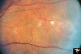 |
Neurofibromatosis-1 | Retinal microvascular malformations in NF-1. Fundus picture shows a somewhat larger vertically running corkscrew malformation between two temporal retinal veins. Pair with R1_E5a. Anatomy: Retina. Pathology: Retinal microvascular malformations. Disease/Diagnosis: Neurofibromatosis type 1. Clinical: ... | Image |
| 6 |
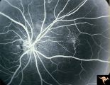 |
Neurofibromatosis-1 | Fluorescein angiogram defines the extent of the microvascular malformation. Pair with R1_E5b. (Ref: BJO 2002:86, p282-284). Anatomy: Retina. Pathology: Retinal microvascular malformations. Disease/Diagnosis: Neurofibromatosis type 1. Clinical: No visual symptoms. Imaging: Fluorescein angiogram. | Image |
| 7 |
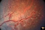 |
Neurofibromatosis-1 | Extensive retinal microvascular malformation involving both small and large retinal vessels. (Ref: BJO 2002:86, p282-284). Anatomy: Retina. Pathology: Retinal microvascular malformations. Disease/Diagnosis: Neurofibromatosis type 1. Clinical: No visual symptoms. | Image |
| 8 |
 |
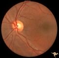
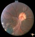
Neurofibromatosis-2 | This is the ocular fundus in a patient with NF-2 showing a preretinal membrane that extends from the temporal disc margin toward the macula. The optic disc shows low grade papilledema caused by one of the patient's acoustic neurinomas. The membrane has caused horizontal folds on the retinal surface.... | Image |
| 9 |
 |
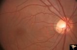
Neurofibromatosis-2 | Retinal tumor in NF-2 referred to as a CPERH (choroidal pigment epithelial retinal hamartoma). Patient, a 16 year old girl, had bilateral acoustic neurinomas. Pair with R1_F2b. Same eye. Anatomy: Optic disc; Retina. Pathology: Retinal hamartoma; Bilateral acoustic neurinoma. Disease/Diagnosis: Neuro... | Image |
| 10 |
 |
Neurofibromatosis-2 | Retinal tumor in NF-2 referred to as a CPERH (choroidal pigment epithelial retinal hamartoma). Patient, a 16 year old girl, had bilateral acoustic neurinomas. Pair with R1_F2a. Same eye. Anatomy: Optic disc; Retina. Pathology: Retinal hamartoma; Bilateral acoustic neurinoma. Disease/Diagnosis: Neuro... | Image |
| 11 |
 |
Neurofibromatosis-2 | CPERH (choroidal pigment epithelial retinal hamartoma) lesion in a patient with NF-2. Note the oblique superficial retinal traction folds running toward the center of the main lesion. 51 year old man. Anatomy: Retina. Pathology: Hamartoma. Disease/Diagnosis: Neurofibromatosis type 2. Clinical: Fiel... | Image |
1 - 25 of 11