Best known for his world-renowned neuro-ophthalmology unit based at the University of California, San Francisco, William Hoyt, MD collected here more than 850 of his best images covering a wide range of disorders.
William F. Hoyt, MD, Professor Emeritus of Ophthalmology, Neurology and Neurosurgery, Department of Ophthalmology, University of California, San Francisco.
NOVEL: https://novel.utah.edu/
TO
Filters: Collection: "ehsl_novel_wfh"
1 - 25 of 16
| Title | Description | Type | ||
|---|---|---|---|---|
| 1 |
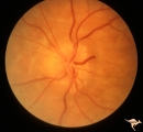 |
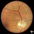
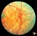
F201 Optic Nerve Sheath Meningioma | Right eye. Woman with ophthalmoplegia proptosis for 14 years. Visual field reduced due to optic nerve sheath meningioma. Notice large optociliary vessel temporally. Anatomy: Optic disc. Pathology: Chronic optic disc swelling caused by optic nerve sheath meningioma. Disease/ Diagnosis: Chronic optic ... | Image |
| 2 |
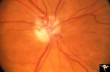 |
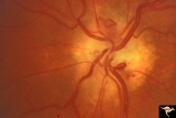
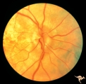
IA01 Atrophy with Optociliary Veins | 1994, perioptic nerve sheath meningioma, right eye, Optociliary vein dumping into disc edge at 4:00. Anatomy: Optic disc. Pathology: Optociliary vein. Disease/ Diagnosis: Perioptic nerve sheath meningioma. Clinical: Progressive visual loss | Image |
| 3 |
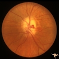 |
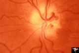
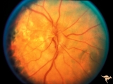
IA02 Atrophy with Optociliary Veins | 1971, left eye, perioptic nerve sheath meningioma, notice how vein dumps into adjacent choroid at 3:00. The darker venous blood can be seen at the disc edge. Anatomy: Optic disc. Pathology: Optociliary vein. Disease/ Diagnosis: Perioptic nerve sheath meningioma. Clinical: Progressive visual loss. | Image |
| 4 |
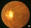 |
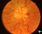
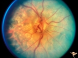
IA03 Atrophy with Optociliary Veins | 1974, left eye, perioptic nerve sheath meningioma, blind eye. Optociliary bypass veins in the nasal disc tissue. Anatomy: Optic disc. Pathology: Optociliary vein. DIsease/ Diagnosis: Perioptic nerve sheath meningioma. Clinical: Blind eye. | Image |
| 5 |
 |
IA04 Atrophy with Optociliary Veins | 1981, right eye, perioptic nerve sheath meningioma with optociliary bypass vein. Notice horizontal choroidal folds in the retina from posterior tumor pressure. Anatomy: Optic disc. Pathology: Optociliary vein. Disease/ Diagnosis: Perioptic nerve sheath meningioma. Clinical: Blind eye. | Image |
| 6 |
 |
IA05 Atrophy with Optociliary Veins | 1971, right eye, perioptic nerve sheath meningioma with optociliary bypass veins on the upper half of the disc. Anatomy: Optic disc. Pathology: Optociliary vein. Disease/ Diagnosis: Perioptic nerve sheath meningioma. Clinical: Blind eye. | Image |
| 7 |
 |
IA06 Atrophy with Optociliary Veins | 1979, left eye, perioptic nerve sheath meningioma with optociliary bypass veins. Anatomy: Optic disc. Pathology: Optociliary vein. Disease/ Diagnosis: Perioptic nerve sheath meningioma. Clinical: Blind eye. | Image |
| 8 |
 |
IA07 Atrophy with Optociliary Veins | Left eye, perioptic nerve sheath meningioma. Anatomy: Optic disc. Pathology: Optociliary vein. Disease/ Diagnosis: Perioptic nerve sheath meningioma. Clinical: Visual loss. | Image |
| 9 |
 |
IA08 Atrophy with Optociliary Veins | 1996, left eye. Chronic pale optic nerve swelling with optociliary bypass veins produced by perioptic nerve sheath meningioma. Anatomy: Optic disc. Pathology: Optociliary vein. Disease/ Diagnosis: Perioptic nerve sheath meningioma evolution. Clinical: Visual loss. | Image |
| 10 |
 |
IA09a Evolution of Optociliary Veins with Perioptic Nerve Sheath Meningioma | April 1975, Normal eye, macular degeneration. Anatomy: Optic disc. Pathology: Optociliary vein. Disease/ Diagnosis: Perioptic nerve sheath meningioma evolution. Clinical: Visual loss. | Image |
| 11 |
 |
IA09b Evolution of Optociliary Veins with Perioptic Nerve Sheath Meningioma | January 1977, macular degeneration, disc swelling begins. Anatomy: Optic disc. Pathology: Optociliary vein. Disease/ Diagnosis: Perioptic nerve sheath meningioma evolution. Clinical: Visual loss. | Image |
| 12 |
 |
IA09c Evolution of Optociliary Veins with Perioptic Nerve Sheath Meningioma | June 1977, continued disc swelling. Anatomy: Optic disc. Pathology: Optociliary vein. Disease/ Diagnosis: Perioptic nerve sheath meningioma evolution. Clinical: Visual loss. | Image |
| 13 |
 |
IA09d Evolution of Optociliary Veins with Perioptic Nerve Sheath Meningioma | October 1977, continued disc swelling. Anatomy: Optic disc. Pathology: Optociliary vein. Disease/ Diagnosis: Perioptic nerve sheath meningioma evolution. Clinical: Visual loss. | Image |
| 14 |
 |
IA09e Evolution of Optociliary Veins with Perioptic Nerve Sheath Meningioma | February 1979, development of optociliary veins at 7:00, 1:00. Anatomy: Optic disc. Pathology: Optociliary vein. Disease/ Diagnosis: Perioptic nerve sheath meningioma evolution. Clinical: Visual loss. | Image |
| 15 |
 |
IA09f Evolution of Optociliary Veins with Perioptic Nerve Sheath Meningioma | August 1979, less disc swelling and development of atrophy with more prominent optociliary veins at 7:00 and 1:00. Anatomy: Optic disc. Pathology: Optociliary vein. Disease/ Diagnosis: Perioptic nerve sheath meningioma evolution. Clinical: Visual loss. | Image |
| 16 |
 |
IA09g Evolution of Optociliary Veins with Perioptic Nerve Sheath Meningioma | April 1980, prominent atrophy and increased numbers of optociliary veins. Anatomy: Optic disc. Pathology: Optociliary vein. Disease/ Diagnosis: Perioptic nerve sheath meningioma evolution. Clinical: Visual loss. | Image |
1 - 25 of 16