Best known for his world-renowned neuro-ophthalmology unit based at the University of California, San Francisco, William Hoyt, MD collected here more than 850 of his best images covering a wide range of disorders.
William F. Hoyt, MD, Professor Emeritus of Ophthalmology, Neurology and Neurosurgery, Department of Ophthalmology, University of California, San Francisco.
NOVEL: https://novel.utah.edu/
Filters: Collection: "ehsl_novel_wfh"
1 - 25 of 15
| Title | Description | Type | ||
|---|---|---|---|---|
| 1 |
 |
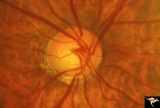
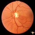
IF101 Low Tension Glaucoma | Low tension glaucoma. Highly myopic eye with shallow cup. Peripapillary choroidal pigment atrophy. Note the narrowed retinal arterioles. 1965. Anatomy: Optic disc. Pathology: Glaucoma. Disease/ Diagnosis: Low tension glaucoma. Clinical: Visual field defects. | Image |
| 2 |
 |
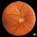
IF102a Low Tension Glaucoma | Low tension glaucoma with bilateral superior altitudinal field defects. Thinning of the neuroretinal rim. Cupping predominantly inferiorly. Pair with IF1_2b. Anatomy: Optic disc. Pathologhy: Glaucoma. Disease/ Diagnosis: Low tension glaucoma. Clinical: Bilateral altitudinal visual field loss. | Image |
| 3 |
 |
IF102b Low Tension Glaucoma | Low tension glaucoma with bilateral superior altitudinal field defects. Thinning of the neuroretinal rim. Cupping predominantly inferiorly. Pair with IF1_2b. Anatomy: Optic disc. Pathology: Glaucoma. Disease/ Diagnosis: Low tension glaucoma. Clinical: Bilateral altitudinal visual field loss. | Image |
| 4 |
 |
IF103a Low Tension Glaucoma | 60 year old woman. Congenital myopia. Temporal pallor. Shallow cupping. Possible low tension glaucoma. Pair with IF1_3b. Note arteriola narrowing. 1971. Anatomy: Optic disc. Clinical: Bilateral field defects. | Image |
| 5 |
 |
IF103b Low Tension Glaucoma | 60 year old woman. Congenital myopia. Temporal pallor. Shallow cupping. Possible low tension glaucoma. Pair with IF1_3a. Note arteriola narrowing. Anatomy: Optic disc. Clinical: Bilateral field defects. | Image |
| 6 |
 |
IF104a Low Tension Glaucoma | Possible low tension glaucoma. Patient with macro discs with remarkable cupping. Pair with IF1_4b. 1969. Anatomy: Optic disc. Disease/ Diagnosis: Cupping and megalopapilla (macrodisc). Clinical: Possible visual field defect. | Image |
| 7 |
 |
IF104b Low Tension Glaucoma | Possible low tension glaucoma. Patient with macro discs with remarkable cupping. Pair with IF1_4a. 1969. Anatomy: Optic disc. Disease/ Diagnosis: Cupping and megalopapilla (macrodisc). Clinical: Possible visual field defect. | Image |
| 8 |
 |
IF105a Low Tension Glaucoma | 40 year old man. Megalopapilla. Right eye has superior arcuate field defect. Pair with IF1_5b. Anatomy: Optic disc. Disease/ Diagnosis: Cupping and megalopapilla (macrodisc). Clinical: Asymptomatic. | Image |
| 9 |
 |
IF105b Low Tension Glaucoma | 40 year old man. Megalopapilla. Left eye. Pair with IF1_5a. Anatomy: Optic disc. Disease/ Diagnosis: Cupping and megalopapilla (macrodisc). Clinical: Asymptomatic. | Image |
| 10 |
 |
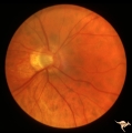
IF106 Low Tension Glaucoma | Low tension glaucoma with subtle inferior temporal wedge defect in the retinal nerve fiber layer corresponding with an inferior temporal defect in the neuroglial rim. 27 year old man. 1984. Anatomy: Optic disc. Pathology: Glaucoma. Disease/ Diagnosis: Low tension glaucoma. Clinical: Superior arcuate... | Image |
| 11 |
 |
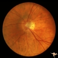
IF110 Low Tension Glaucoma | Low tension glaucoma with an inferior sector defect in the retinal nerve fiber layer. 1979. Anatomy: Optic disc. Pathology: Glaucoma. Disease/ Diagnosis: Low tension glaucoma. Clinical: Superior field defects. | Image |
| 12 |
 |
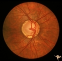
IF111a Low Tension Glaucoma | Low tension glaucoma. Followed. Pair with IF1_11b, c, d. Left eye. 1981. Anatomy: Optic disc. Pathology: Glaucoma. Disease/ Diagnosis: Low tension glaucoma. Clinical: Asymptomatic. | Image |
| 13 |
 |
IF111b Low Tension Glaucoma | Low tension glaucoma. Followed, 9 years later. Wedge defects in retinal nerve fiber defects in both temporal arcuate zones. Note small disc edge hemorrhage at 5:00. Pair with IF1_11a, c, d. Left eye. 1990. Anatomy: Optic disc. Pathology: Glaucoma. Disease/ Diagnosis: Low tension glaucoma. Clinical:... | Image |
| 14 |
 |
IF111c Low Tension Glaucoma | Low tension glaucoma. Followed. Notice disc edge hemorrhage at 7:00. Inferior nerve fiber layer defect between 6:00 and 7:30.Pair with IF1_11a, b, d. Right eye. 1981. Anatomy: Optic disc. Pathology: Glaucoma. Disease/ Diagnosis: Low tension glaucoma. Clinical: Superior arcuate visual field defect | Image |
| 15 |
 |
IF111d Low Tension Glaucoma | Low tension glaucoma. Followed. Inferior arcuate field defect has expanded upward. Note increase in atrophy and cupping in inferior temporal disc. Pair with IF1_11a, b, d. Right eye. 1990. Anatomy: Optic disc. Pathology: Glaucoma. Disease/ Diagnosis: Low tension glaucoma. Clinical: Increased size o... | Image |
1 - 25 of 15