Best known for his world-renowned neuro-ophthalmology unit based at the University of California, San Francisco, William Hoyt, MD collected here more than 850 of his best images covering a wide range of disorders.
William F. Hoyt, MD, Professor Emeritus of Ophthalmology, Neurology and Neurosurgery, Department of Ophthalmology, University of California, San Francisco.
NOVEL: https://novel.utah.edu/
TO
Filters: Collection: "ehsl_novel_wfh"
| Title | Description | Type | ||
|---|---|---|---|---|
| 1 |
 |
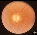
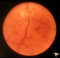
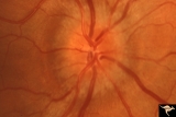
Bilateral Papilledema with Cyanotic Heart Disease | Bilateral Papilledema with cyanotic heart disease in a young boy. Anatomy: Optic disc. Pathology: Papilledema. Disease/Diagnosis: Pseudotumor due to cyanotic heart disease. Clinical notes: Young boy with clubbing. | Image |
| 2 |
 |
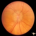
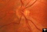
C38 Anomalous Pale Disc | Megalopapilla in -8 myopic eye. Right eye. Anatomy: Optic disc. Clinical: High myope. | Image |
| 3 |
 |
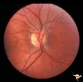
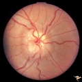
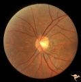
H19 Panhypoplasia | Mild hypoplasia with dysplasia in right eye. Right eye. Normal left eye. Same patient as H_20. Anatomy: Optic disc. Pathology: Hypoplasia of the optic nerve. Disease/ Diagnosis: Hypoplasia. | Image |
| 4 |
 |
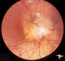
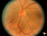
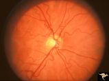
Multifocal Choroidopathy | Multifocal choroidopathy in a patient with uveitis. Anatomy: Retina. Disease/Diagnosis: Acute Multifocal Placoid Pigment Epitheliopathy (AMPPE). Clinical: Visual loss. | Image |
| 5 |
 |
Tuberous Sclerosis | Tuberous Sclerosis. Astrocytic hamartoma of the optic disc. Anatomy: Optic disc. Pathology: Astrocytic hamartoma. Disease/Diagnosis: Tuberous sclerosis. Clinical: No visual symptoms. | Image |
| 6 |
 |
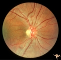
Unilateral Papilledema | Right eye. Has no optic cup. Patient has pseudotumor cerebri. Woman. Anatomy: Optic disc. Pathology: Unilateral papilledema. Disease/Diagnosis: Idiopathic intracranial hypertension, pseudotumor cerebri. Clinical: Woman, headache, transient visual obscurations. | Image |
| 7 |
 |
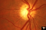
Chronic Papilledema with Pseudo Drusen | Left eye. Meningioma. Pseudo drusen from chronic papilledema. The patient's meningioma had blinded her left eye and caused chronic elevated intracranial pressure. Woman. Anatomy: Optic disc Pathology: Papilledema Disease/Diagnosis: Chronic papilledema with pseudo drusen | Image |
| 8 |
 |
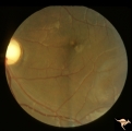
F101 Optic Disc Lymphosarcoma | Optic disc lymphosarcoma. This disc has been infiltrated by neoplastic cells. Anatomy: Optic disc. Pathology: Lymphosarcoma. Disease/ Diagnosis: Lymphosarcoma. | Image |
| 9 |
 |
IA08 Atrophy with Optociliary Veins | 1996, left eye. Chronic pale optic nerve swelling with optociliary bypass veins produced by perioptic nerve sheath meningioma. Anatomy: Optic disc. Pathology: Optociliary vein. Disease/ Diagnosis: Perioptic nerve sheath meningioma evolution. Clinical: Visual loss. | Image |
| 10 |
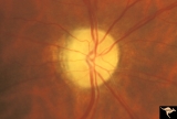 |
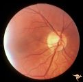
IB104 Post Ischemic (AION) Cupless Atrophy | January 1969, right eye, striking focal arteriolar narrowing at 4:00 and 10:00. Anatomy: Optic disc. Pathology: Post ischemic (AION) cupless atrophy. Disease/ Diagnosis: Post ischemic (AION) cupless atrophy. Clinical: Visual loss. | Image |
| 11 |
 |
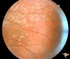
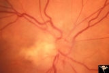
Slow Flow (Chronic Hypoxic) Retinopathy | Slow flow (chronic hypoxic) retinopathy from macroglobulanemia. Note the dot and blot hemorrhages. Anatomy: Retina. Pathology: Macroglobulanemia. Disease/Diagnosis: Slow flow (chronic hypoxic) retinopathy from macroglobulanemia. | Image |
| 12 |
 |
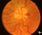
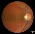
Unilateral Papilledema | Unilateral papilledema in Pseudotumor cerebri in patient with elevated intracranial pressure. Right eye. Anatomy: Optic disc. Pathology: Unilateral papilledema. Disease/Diagnosis: Idiopathic intracranial hypertension (pseudotumor cerebri). Clinical: Transient monocular blindness (transient visual ob... | Image |
| 13 |
 |
Visible Drusen - Bilateral | PP22a: right eye with obvious exposed drusen. PP22 b: Note bypass vein draining into the choroid at 8:00. Anatomy: Optic disc. Pathology: Drusen of the optic disc. Disease/Diagnosis: Drusen of the optic disc. Clinical: Normally functioning eye with drusen. | Image |
| 14 |
 |
Von Hippel Lindau Disease (Retinal Hemangioblastoma) | Von Hippel Lindau Disease (Retinal Hemangioblastoma); Small hemangioblastoma on the disc margin at 10:00. Large peripheral hemangioblastoma out of view to the top right seen on R1_C2b. Anatomy: Optic disc. Pathology: Hemangioblastoma. Disease/Diagnosis: Von Hippel Lindau disease. Clinical: No visual... | Image |
| 15 |
 |
C105 Disc Edema with Systemic Lupus | Mild disc edema blurs the inferior disc margin. Flourescein angiogram in D1_06. Same patient as D1_06 an D1_07. Man. Anatomy: Optic disc. Pathology: Axoplasmic stasis due to vasculitis (Lupus). Disease/ Diagnosis: Lupus papillopathy. | Image |
| 16 |
 |
D103 Disc Edema with Systemic Lupus | 28 year old woman with systemic Lupus erythematosus. Vision 20/20 but blind spot enlarged. Same patient as D1_02. Magnified. Anatomy: Optic disc. Pathology: Axoplasmic stasis due to vasculitis (Lupus). Disease/ Diagnosis: Lupus papillopathy. Clinical: Normal vision with enlarged blind spot on visual... | Image |
| 17 |
 |
H34 Segmental Hypoplasia, Retinal-Congenital Toxo | Left eye. Temporal sector hypoplasia from congenital retinal toxoplasmosis. Note the sector shaped nerve fiber loss between 2:00 and 4:00. Same patient as H_35. Anatomy: Optic disc; retina. Pathology: Hypoplasia secondary to retinal lesion. Disease/ Diagnosis: Segmental optic disc hypoplasia. | Image |
| 18 |
 |
H43 Segmental Hypoplasia, Retinal, Nasal Hypoplasia | Nasal hypoplasia barely perceptible on disc. Nasal retinal nerve fibers are completely absent from 7:00 - 12:00. Anaotmy: Optic disc. Pathology: Nasal segmental disc hypoplasia. Disease/ Diagnosis: Congenital anomaly. | Image |
| 19 |
 |
H73 Superior Segmental Optic Hypoplasia (SSOH) Topless Disc Syndrome | Bilateral SSOH. Left eye. Same patient as H_73. Anatomy: Optic disc. Pathology: Superior segmental optic hypoplasia (SSOH). Disease/ Diagnosis: Congenital anomaly. | Image |
| 20 |
 |
IE08a Subacute Leber Optic Neuropathy | Subacute Leber Optic Neuropathy with temporal atrophy. August 5, 1980. Pair with IE_1, 2a&b, IE_8b, IE_9a&b. Anatomy: Optic disc. Pathology: Optic neuropathy. Disease/ Diagnosis: Leber's optic neuropathy. Clinical: Visual loss. | Image |
| 21 |
 |
IE08b Subacute Leber Optic Neuropathy | Subacute Leber Optic Neuropathy with temporal atrophy. August,1980. Pair with IE_1, 2a&b, IE_8a, IE_9a&b. Anatomy: Optic disc. Pathology: Optic neuropathy. Disease/ Diagnosis: Leber's optic neuropathy. Clinical: Visual loss. | Image |
| 22 |
 |
IIA102a Diffuse Atrophy | Bilateral primary or retrograde optic atrophy from bilateral optic nerve sheath meningiomas. Pair with IIA1_2b. Right eye. 1984. Anatomy: Optic disc. Pathology: Bilateral optic nerve sheath meningiomas. Disease/ Diagnosis: Retrograde optic atrophy. Clinical: Bilateral visual loss. | Image |
| 23 |
 |
Segmental Atrophy - Temporal | Segmental Atrophy - Temporal - Nutritional amblyopia (alcoholic). 1985. Left eye. Pair with IIA2_03a. Anatomy: Optic disc.. Pathology: Optic atrophy. Disease/Diagnosis: Toxic optic atrophy from alcohol. Clinical: Central visual loss. | Image |
| 24 |
 |
Diffuse Atrophy | Nerve fiber appearance about 6 weeks after indirect injury to optic nerve. Note near total absence of nerve fiber reflexes. Photo shows remaining streaks of inferior arcuate nerve fiber membranes dissolving into nothing. 1972. Anatomy: Optic disc. Pathology: Optic nerve injury. Disease/Diagnosis: Op... | Image |
| 25 |
 |
IE09a Chronic Leber Optic Neuropathy | Chronic Leber Optic Neuropathy with advancing temporal pallor. Notice the nerve fiber layer thickening has diminished. November 13, 1980. Pair with IE_1, 2a&b, IE_9b, IE_8a&b. Anatomy: Optic disc. Pathology: Optic neuropathy. Disease/ Diagnosis: Leber's optic neuropathy. Clinical: Blindness. | Image |