Best known for his world-renowned neuro-ophthalmology unit based at the University of California, San Francisco, William Hoyt, MD collected here more than 850 of his best images covering a wide range of disorders.
William F. Hoyt, MD, Professor Emeritus of Ophthalmology, Neurology and Neurosurgery, Department of Ophthalmology, University of California, San Francisco.
NOVEL: https://novel.utah.edu/
TO
Filters: Collection: "ehsl_novel_wfh"
| Title | Description | Type | ||
|---|---|---|---|---|
| 1 |
 |
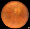
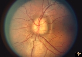
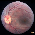
Ischemic Complication of Drusen | PP30a: right eye--buried drusen; PP30b: buried drusen with anterior ischemic optic neuropathy (AION) from complication of drusen of left eye. Ischemic complication of drusen in left eye. PP30c: 3 month follow-up: narrowed arterioles slightly pale disc with buried drusen. Anatomy: Optic disc. Patho... | Image |
| 2 |
 |
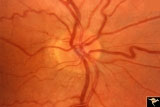
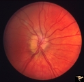
Visible Drusen | PP21a: Right eye. Drusen barely visible. Note disc margin drusen at 1:00 and 2:30.; PP21b: Left eye shows multiple exposed drusen. Girl. Anatomy: Optic disc. Pathology: Drusen of the optic disc. Disease/Diagnosis: Drusen of the optic disc. Clinical: Normally functioning eye with drusen. | Image |
| 3 |
 |
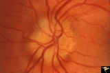
Ischemic Complication of Drusen | PP30a: right eye--buried drusen; PP30b: buried drusen with anterior ischemic optic neuropathy (AION) from complication of drusen of left eye. Ischemic complication of drusen in left eye. PP30c: 3 month follow-up: narrowed arterioles slightly pale disc with buried drusen. Anatomy: Optic disc. Patho... | Image |
| 4 |
 |
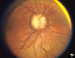
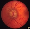
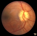
Familial Drusen | Right eye: Mother with obvious optic nerve drusen. Note the blurred temporal margin where buried drusen can not be seen.; PP_11b: mother visible drusen; Buried drusen; lumpy disc. Combine with PP_1a & b and PP_2 (sons) and PP_11c (daughter). Anatomy: Optic disc. Pathology: Drusen of the optic dis... | Image |
| 5 |
 |
Ischemic Complication of Drusen | PP30a: right eye--buried drusen; PP3-b: buried drusen with anterior ischemic optic neuropathy (AION) from complication of drusen of left eye. Ischemic complication of drusen in left eye. PP30c: 3 month follow-up: narrowed arterioles slightly pale disc with buried drusen. Anatomy: Optic disc. Path... | Image |
| 6 |
 |
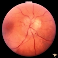
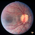
Unilateral Buried Drusen | PP20a: Right eye. Normal disc without optic cup.PP20b: Left eye. Buried drusen nasally and exposed drusen at the temporal margin. Boy. Anatomy: Optic disc. Pathology: Drusen of the optic disc. Disease/Diagnosis: Drusen of the optic disc. Clinical: Normally functioning eye with drusen. Right eye nor... | Image |
| 7 |
 |
Unilateral Buried Drusen | PP20a: Right eye. Normal disc without optic cup.PP20b: Left eye. Buried drusen nasally and exposed drusen at the temporal margin. Boy. Anatomy: Optic disc. Pathology: Drusen of the optic disc.. Disease/Diagnosis: Drusen of the optic disc.. Clinical: Normally functioning eye with drusen. Right eye n... | Image |
| 8 |
 |
Familial Drusen | Left eye. PP_11b: Mother visible drusen; buried drusen; lumpy disc. PP_11a: Mother with obvious optic nerve drusen; Combine with PP_1a & b and PP_2 (sons) and PP_11c (daughter). Anatomy: Optic disc. Pathology: Drusen of the optic disc. Disease/Diagnosis: Drusen of the optic disc. Clinical: Congen... | Image |
| 9 |
 |
Buried and Visible Drusen | PP_19b: right eye : visible drusen in an eleven year old girl; PP_19a: left eye with buried drusen. Anatomy: Optic disc Pathology: Drusen of the optic disc Disease/Diagnosis: Drusen of the optic disc Clinical: Normally functioning eye with drusen. | Image |
| 10 |
 |
Buried and Visible Drusen | PP_19a Left eye with buried drusen. PP_19b: right eye : visible drusen. Eleven year old girl. Anatomy: Optic disc. Pathology: Drusen of the optic disc. Disease/Diagnosis: Drusen of the optic disc. Clinical notes: Normally functioning eye with drusen. | Image |
| 11 |
 |
Buried Drusen | Buried drusen with peculiar white dot, which appears to be choroidal in location. Note lumpy disc margin on right disc PP_15a is right eye. PP_15b is left eye. Beautiful example of pseudo papilledema in which drusen can not be seen. 8 year old girl. Anatomy: Optic disc. Pathology: Drusen of the op... | Image |
| 12 |
 |
Buried Drusen | Buried drusen; PP_13a: Right eye. Note lumpy disc margin, especially temporally. Also note absence of optic cup. Excellent example of pseudo papilledema with buried drusen. Anatomy: Optic disc. Pathology: Drusen of the optic disc. Disease/Diagnosis: Drusen of the optic disc. Clinical notes: Patient ... | Image |
| 13 |
 |
Buried Drusen | Buried drusen. Left eye. Note lumpy disc margin, especially temporally. Also note absence of optic cup. Excellent example of pseudo papilledema with buried drusen. Pair with PP_13a. Anatomy: Optic disc. Pathology: Drusen of the optic disc. Disease/Diagnosis: Drusen of the optic disc. Clinical notes... | Image |
| 14 |
 |
Drusen Plus Papilledema | PP37a: right swollen disc on top of drusen with narrowing of the arterioles; PP37b: left visible drusen and papilledema with sub-retinal hemorrhage temporally. Patient had frontal glioblastoma. Anatomy: Optic disc. Pathology: Drusen of the optic disc. Disease/Diagnosis: Drusen of the optic disc. Cli... | Image |
| 15 |
 |
Visible Drusen with Visual Field Loss | Left eye.16 year old girl: PP26b: buried drusen at the lower pole of the disc; PP26a: Visible drusen with visual field loss. Notice the thinning of the nerve fibers in both the superior and inferior arcuate bundles. PP26c: Goldmann visual field. Anatomy: Optic disc. Pathology: Drusen of the optic ... | Image |
| 16 |
 |
Visible Drusen with Visual Field Loss | Right eye.16 year old girl: PP26a: Visible drusen with visual field loss. Notice the thinning of the nerve fibers in both the superior and inferior arcuate bundles. PP26b: buried drusen; PP26c: Goldmann visual field. Anatomy: Optic disc. Pathology: Drusen of the optic disc. Disease/Diagnosis: Druse... | Image |
| 17 |
 |
Buried Drusen | 5 year old boy. Bilateral buried drusen. Notice the lumpy nasal disc elevation. This patient had a twin brother whose optic disc drusen were exposed. Anatomy: Optic disc. Pathology: Drusen of the optic disc. Disease/Diagnosis: Drusen of the optic disc. Clinical notes: Normally functioning eye with ... | Image |
| 18 |
 |
Buried Drusen | 5 year old boy. Bilateral buried drusen. Notice the lumpy nasal disc elevation. This patient had a twin brother whose optic disc drusen were exposed. Anatomy: Optic disc. Pathology: Drusen of the optic disc. Disease/Diagnosis: Drusen of the optic disc. Clinical notes: Normally functioning eye with ... | Image |
| 19 |
 |
Buried Drusen | Suspected buried drusen in a girl. Anatomy: Optic disc. Pathology: Drusen of the optic disc. Disease/Diagnosis: Drusen of the optic disc. Clinical notes: Normally functioning eye with suspected drusen. | Image |
| 20 |
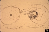 |
Visible Drusen with Visual Field Loss | 16 year old girl: Drusen disc. Goldmann visual field. Anatomy: Optic disc. Pathology: Drusen of the optic disc. Disease/Diagnosis: Drusen of the optic disc. Clinical: Drusen disc with visual field loss. | Image |
| 21 |
 |
Vascular Complications of Drusen: Drusen Causing Loss of Superior Retinal Arterial Supply | PP32a: right; PP32b: left eye. Right eye is an obvious drusen disc. Patient had marked field defects. Left eye has occlusion of superior branch of the central retinal artery at 11:30 with the inferior retinal artery supplying collateral to the superior retina. Notice the branch of the inferior ret... | Image |
| 22 |
 |
Buried Drusen | Excellent example of pseudo papilledema with sub surface drusen at 10:00 and 1:00. Anatomy: Optic disc. Pathology: Drusen of the optic disc. Disease/Diagnosis: Drusen of the optic disc. Clinical notes: Normally functioning eye with drusen. | Image |
| 23 |
 |
Drusen with Horizontal Retinal Folds | PP35a: Right eye. Buried drusen. PP35b: Left eye. Buried drusen with retinal folds. 21 year old woman. Anatomy: Optic disc. Pathology: Drusen of the optic disc. Disease/Diagnosis: Drusen of the optic disc. | Image |
| 24 |
 |
Drusen with Horizontal Retinal Folds | PP35: Right eye. Buried drusen. PP35b: Left eye. Buried drusen with retinal folds. 21 year old woman. Anatomy: Optic disc. Pathology: Drusen of the optic disc. Disease/Diagnosis: Drusen of the optic disc. | Image |
| 25 |
 |
Familial Drusen | PP11c: daughter: buried drusen; lumpy disc. Combine with PP1a & b and PP2 (brothers) and PP11a & b (mother). Anatomy: Optic disc. Pathology: Drusen of the optic disc. Disease/Diagnosis: Drusen of the optic disc. Clinical: Congenital dominant hereditary drusen. | Image |