Best known for his world-renowned neuro-ophthalmology unit based at the University of California, San Francisco, William Hoyt, MD collected here more than 850 of his best images covering a wide range of disorders.
William F. Hoyt, MD, Professor Emeritus of Ophthalmology, Neurology and Neurosurgery, Department of Ophthalmology, University of California, San Francisco.
NOVEL: https://novel.utah.edu/
TO
| Title | Description | Type | ||
|---|---|---|---|---|
| 126 |
 |
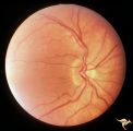
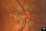
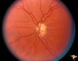
H105 Occipital Hemianoptic Hypoplasia | Left congenital homonymous hemianopia. Right occipital AVM. Nasal nerve fiber layer loss in left eye. Compare with right eye. Same patient as H_106. Anatomy: Optic disc. Pathology: Occipital hemianoptic hypoplasia. DIsease/ Diagnosis: Congenital defect of the occipital lobe | Image |
| 127 |
 |
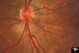
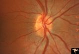
H106 Occipital Hemianoptic Hypoplasia | Same patient as H_105. Anatomy: Optic disc. Pathology: Occipital hemianoptic hypoplasia. Disease/ Diagnosis: Congenital defect of the occipital lobe. | Image |
| 128 |
 |
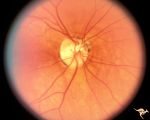
H15 Panhypoplasia | Moderate hypoplasia. Right eye. 14 year old boy. Good example of double ring sign. Same patient as H_16. Anatomy: Optic disc. Pathology: Hypoplasia of the optic nerve. Disease/ Diagnosis: Hypoplasia. | Image |
| 129 |
 |
H16 Panhypoplasia | Moderate hypoplasia. Left eye. 14 year old boy. Good example of double ring sign. Same patient as H_15. Anatomy: Optic disc. Pathology: Hypoplasia of the optic nerve. Disease/ Diagnosis: Hypoplasia. | Image |
| 130 |
 |
H17 Panhypoplasia | Bilateral mild hypoplasia without field defect. Right eye. 30 year old woman. Same patient as H_18. Anatomy: Optic disc. Pathology: Hypoplasia of the optic nerve. Disease/ Diagnosis: Hypoplasia. | Image |
| 131 |
 |
H18 Panhypoplasia | Bilateral mild hypoplasia without field defect. Left eye. 30 year old woman. Same patient as H_17. Anatomy: Optic disc. Pathology: Hypoplasia of the optic nerve. Disease/ Diagnosis: Hypoplasia. | Image |
| 132 |
 |
H19 Panhypoplasia | Mild hypoplasia with dysplasia in right eye. Right eye. Normal left eye. Same patient as H_20. Anatomy: Optic disc. Pathology: Hypoplasia of the optic nerve. Disease/ Diagnosis: Hypoplasia. | Image |
| 133 |
 |
H20 Panhypoplasia | Mild hypoplasia with dysplasia in right eye. Left eye. Same patient as H_19. Anatomy: Optic disc. Pathology: Hypoplasia of the optic nerve. Disease/ Diagnosis: Hypoplasia. | Image |
| 134 |
 |
H36 Segmental Hypoplasia, Retinal, Congenital Toxo | Left eye. Optic disc hypoplasia from congenital nasal retinal toxoplasma lesion. Chorioretinal scar. Anatomy: Optic disc, retina. Pathology: Hypoplasia secondary to retinal lesion. Disease/ Diagnosis: Segmental optic disc hypoplasia. | Image |
| 135 |
 |
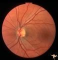
H40 Segmental Hypoplasia, Retinal, Tilted (Dysverted) Disc | 60 year old woman with incidental bitemporal visual field depression. Extreme tilting of optic disc with inferior nasal segmental hypoplasia. Nasal retinal ectasia. Same patient as H_41. Anatomy: Optic disc; retina. Pathology: Hypoplasia secondary to retinal lesion. Disease/ Diagnosis: Segmental opt... | Image |
| 136 |
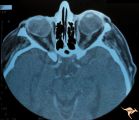 |
H41 Segmental Hypoplasia, Retinal, Tilted (Dysverted) Disc | CT scan of patient in H_40 showing marked nasal ectasia of the eyeballs. CT scan shows obliquely inserted optic nerves and marked nasal dysplasia of the eyeballs. Anatomy: Optic disc; retina. Pathology: Hypoplasia secondary to retinal lesion. Disease/ Diagnosis: Segmental optic disc hypoplasia. Imag... | Image |
| 137 |
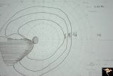 |
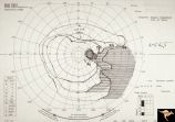
H42 Segmental Hypoplasia, Retinal, Nasal Hypoplasia | Visual field of patient in H_43. Flag-like temporal field defect from patient with nasal segmental disc hypoplasia. Anatomy: Optic disc. Pathology: Nasal segmental disc hypoplasia. Disease/ Diagnosis: Congenital anomaly. | Image |
| 138 |
 |
H43 Segmental Hypoplasia, Retinal, Nasal Hypoplasia | Nasal hypoplasia barely perceptible on disc. Nasal retinal nerve fibers are completely absent from 7:00 - 12:00. Anaotmy: Optic disc. Pathology: Nasal segmental disc hypoplasia. Disease/ Diagnosis: Congenital anomaly. | Image |
| 139 |
 |
H44 Segmental Hypoplasia, Retinal, Nasal Hypoplasia | Bilateral nasal hypoplasia with bilateral flag-like temporal field defect. Right eye. Same patient as H_45. Anatomy: Optic disc. Pathology: Nasal segmental disc hypoplasia. Disease/ Diagnosis: Congenital anomaly. | Image |
| 140 |
 |
H45 Segmental Hypoplasia, Retinal-Nasal Hypoplasia | Bilateral nasal hypoplasia with bilateral flag-like temporal field defect. Left eye. Same patient as H_44. Anatomy: Optic disc. Pathology: Nasal segmental disc hypoplasia. Disease/ Diagnosis: Congenital anomaly. | Image |
| 141 |
 |
H48 Segmental Hypoplasia, Retinal-Nasal Hypoplasia | Bilateral nasal hypoplasia with absence of nasal nerve fiber layer and corresponding flag-like temporal field defect. Right eye. Same patient as H_49. Anatomy: Optic disc. Pathology: Nasal segmental disc hypoplasia. Disease/ Diagnosis: Congential anomaly. | Image |
| 142 |
 |
H49 Segmental Hypoplasia, Retinal-Nasal Hypoplasia | Bilateral nasal hypoplasia with absence of nasal nerve fiber layer and corresponding flag-like temporal field defect. Left eye. Same patient as H_48. Anatomy: Optic disc. Pathology: Nasal segmental disc hypoplasia. Disease/ Diagnosis: Congential anomaly. | Image |
| 143 |
 |
H50 Segmental Hypoplasia, Retinal-Nasal Hypoplasia | Right eye. Nasal hypoplasia with nasal pit. Same patient as H_51. Anatomy: Optic disc. Pathology: Nasal segmental disc hypoplasia. Disease/ Diagnosis: Congential anomaly. | Image |
| 144 |
 |
H51 Segmental Hypoplasia, Retinal-Nasal Hypoplasia | Visual field showing flag-like temporal field defect in patient shown in H_50. Anatomy: Optic disc. Pathology: Nasal segmental disc hypoplasia. Disease/ Diagnosis: Cogenital anomaly. | Image |
| 145 |
 |
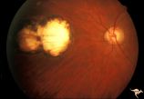
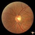
H52 Superior Segmental Optic Hypoplasia (SSOH) Topless Disc Syndrome | High exit point of central retinal vessels. Superior choroidal crescent. Complete loss of nerve fiber layer entering disc from above. Inferior altitudinal field defect. Type 1 diabetic mother. Anatomy: Optic disc. Pathology: Superior segmental optic hypoplasia (SSOH). Disease/ Diagnosis: Congenital ... | Image |
| 146 |
 |
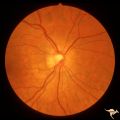
H57 Superior Segmental Optic Hypoplasia (SSOH) Topless Disc Syndrome | Note, in addition to SSOH, generalized hypoplasia of nerve. Anatomy: Optic disc. Pathology: Superior segmental optic hypoplasia (SSOH). Disease/ Diagnosis: Congenital anomaly. | Image |
| 147 |
 |
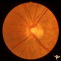
H61 Superior Segmental Optic Hypoplasia (SSOH) Topless Disc Syndrome | Left eye. SSOH. Same patient as H_62 and H_63. Anatomy: Optic disc. Pathology: Superior segmental optic hypoplasia (SSOH). Disease/ Diagnosis: Congenital anomaly. | Image |
| 148 |
 |
H62 Superior Segmental Optic Hypoplasia (SSOH) Topless Disc Syndrome | Right eye. Disc looks almost normal but superior nerve fiber layer is thinned and represents a mild form of SSOH. Same patient as H_61 and H_63. Anatomy: Optic disc. Pathology: Superior segmental optic hypoplasia (SSOH). Disease/ Diagnosis: Congenital anomaly. | Image |
| 149 |
 |
H63 Superior Segmental Optic Hypoplasia (SSOH) Topless Disc Syndrome | Visual fields. Right eye has mild depression. Same patient as H_61 an H_62. Anatomy: Optic disc. Pathology: Superior segmental optic hypoplasia (SSOH). Disease/ Diagnosis: Congenital anomaly. | Image |
| 150 |
 |
H64 Superior Segmental Optic Hypoplasia (SSOH) Topless Disc Syndrome | Bilateral SSOH. Same patient as H_65 and H_66. Anatomy: Optic disc. Pathology: Superior segmental optic hypoplasia (SSOH). Disease/ Diagnosis: Congenital anomaly. | Image |