Best known for his world-renowned neuro-ophthalmology unit based at the University of California, San Francisco, William Hoyt, MD collected here more than 850 of his best images covering a wide range of disorders.
William F. Hoyt, MD, Professor Emeritus of Ophthalmology, Neurology and Neurosurgery, Department of Ophthalmology, University of California, San Francisco.
NOVEL: https://novel.utah.edu/
TO
Filters: Collection: "ehsl_novel_wfh"
| Title | Description | Type | ||
|---|---|---|---|---|
| 501 |
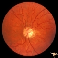 |
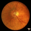
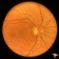
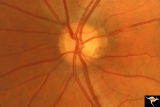
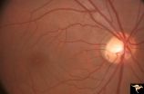
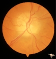
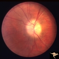
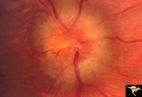
IE14b End Stage Leber Optic Neuropathy | End stage Leber's Optic Neuropathy. Dense temporal pallor. Microangiopathy is absent. Left eye. Pair with 14a. Anatomy: Optic disc. Pathology: Optic neuropathy. Disease/ Diagnosis: Leber's optic neuropathy. Clinical: Blindness. | Image |
| 502 |
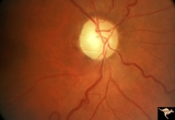 |
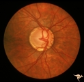
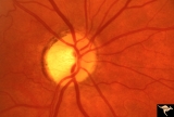
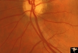
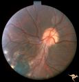
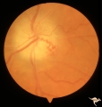
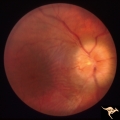
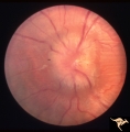
IE15a End Stage Leber Optic Neuropathy | End stage Leber's Optic Neuropathy. Severe diffuse pallor. Right eye. Pair with 15b. Anatomy: Optic disc. Pathology: Optic neuropathy. Disease/ Diagnosis: Leber's optic neuropathy. Clinical: Blindness. | Image |
| 503 |
 |
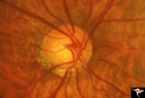
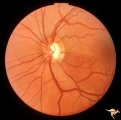
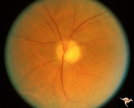
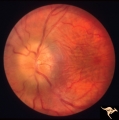
IF101 Low Tension Glaucoma | Low tension glaucoma. Highly myopic eye with shallow cup. Peripapillary choroidal pigment atrophy. Note the narrowed retinal arterioles. 1965. Anatomy: Optic disc. Pathology: Glaucoma. Disease/ Diagnosis: Low tension glaucoma. Clinical: Visual field defects. | Image |
| 504 |
 |
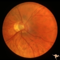
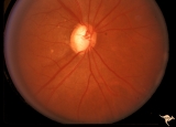
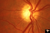
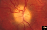
IF102a Low Tension Glaucoma | Low tension glaucoma with bilateral superior altitudinal field defects. Thinning of the neuroretinal rim. Cupping predominantly inferiorly. Pair with IF1_2b. Anatomy: Optic disc. Pathologhy: Glaucoma. Disease/ Diagnosis: Low tension glaucoma. Clinical: Bilateral altitudinal visual field loss. | Image |
| 505 |
 |
IF102b Low Tension Glaucoma | Low tension glaucoma with bilateral superior altitudinal field defects. Thinning of the neuroretinal rim. Cupping predominantly inferiorly. Pair with IF1_2b. Anatomy: Optic disc. Pathology: Glaucoma. Disease/ Diagnosis: Low tension glaucoma. Clinical: Bilateral altitudinal visual field loss. | Image |
| 506 |
 |
IF103a Low Tension Glaucoma | 60 year old woman. Congenital myopia. Temporal pallor. Shallow cupping. Possible low tension glaucoma. Pair with IF1_3b. Note arteriola narrowing. 1971. Anatomy: Optic disc. Clinical: Bilateral field defects. | Image |
| 507 |
 |
IF103b Low Tension Glaucoma | 60 year old woman. Congenital myopia. Temporal pallor. Shallow cupping. Possible low tension glaucoma. Pair with IF1_3a. Note arteriola narrowing. Anatomy: Optic disc. Clinical: Bilateral field defects. | Image |
| 508 |
 |
IF104a Low Tension Glaucoma | Possible low tension glaucoma. Patient with macro discs with remarkable cupping. Pair with IF1_4b. 1969. Anatomy: Optic disc. Disease/ Diagnosis: Cupping and megalopapilla (macrodisc). Clinical: Possible visual field defect. | Image |
| 509 |
 |
IF104b Low Tension Glaucoma | Possible low tension glaucoma. Patient with macro discs with remarkable cupping. Pair with IF1_4a. 1969. Anatomy: Optic disc. Disease/ Diagnosis: Cupping and megalopapilla (macrodisc). Clinical: Possible visual field defect. | Image |
| 510 |
 |
IF105a Low Tension Glaucoma | 40 year old man. Megalopapilla. Right eye has superior arcuate field defect. Pair with IF1_5b. Anatomy: Optic disc. Disease/ Diagnosis: Cupping and megalopapilla (macrodisc). Clinical: Asymptomatic. | Image |
| 511 |
 |
IF105b Low Tension Glaucoma | 40 year old man. Megalopapilla. Left eye. Pair with IF1_5a. Anatomy: Optic disc. Disease/ Diagnosis: Cupping and megalopapilla (macrodisc). Clinical: Asymptomatic. | Image |
| 512 |
 |
IF106 Low Tension Glaucoma | Low tension glaucoma with subtle inferior temporal wedge defect in the retinal nerve fiber layer corresponding with an inferior temporal defect in the neuroglial rim. 27 year old man. 1984. Anatomy: Optic disc. Pathology: Glaucoma. Disease/ Diagnosis: Low tension glaucoma. Clinical: Superior arcuate... | Image |
| 513 |
 |
IF107 Glaucoma Cupped Disc | Glaucoma cupped disc with inferior temporal retinal nerve fiber layer defect. Vertically ovoid cup. 1974. Anatomy: Optic disc. Pathology: Glaucoma. Disease/ Diagnosis: Glaucoma. Clinical: Superior arcuate visual field defects. | Image |
| 514 |
 |
IF108 Glaucoma Cupped Disc | Glaucoma cupped disc. Note dark slits in the upper arcuate retinal nerve fibers. Anatomy: Optic disc. Pathology: Glaucoma. Disease/ Diagnosis: Glaucoma. Clinical: Inferior field defects. | Image |
| 515 |
 |
IF109 Glaucoma Cupped Disc | Glaucoma cupped disc. Note inferior extension of the optic cup, thinning of the neuroglial rim at 5:00 and inferior sector defect in the retinal nerve fiber layer. Anatomy: Optic disc. Pathology: Glaucoma. Disease/ Diagnosis: Glaucoma. Clinical: Superior field defects. | Image |
| 516 |
 |
IF110 Low Tension Glaucoma | Low tension glaucoma with an inferior sector defect in the retinal nerve fiber layer. 1979. Anatomy: Optic disc. Pathology: Glaucoma. Disease/ Diagnosis: Low tension glaucoma. Clinical: Superior field defects. | Image |
| 517 |
 |
IF111a Low Tension Glaucoma | Low tension glaucoma. Followed. Pair with IF1_11b, c, d. Left eye. 1981. Anatomy: Optic disc. Pathology: Glaucoma. Disease/ Diagnosis: Low tension glaucoma. Clinical: Asymptomatic. | Image |
| 518 |
 |
IF111b Low Tension Glaucoma | Low tension glaucoma. Followed, 9 years later. Wedge defects in retinal nerve fiber defects in both temporal arcuate zones. Note small disc edge hemorrhage at 5:00. Pair with IF1_11a, c, d. Left eye. 1990. Anatomy: Optic disc. Pathology: Glaucoma. Disease/ Diagnosis: Low tension glaucoma. Clinical:... | Image |
| 519 |
 |
IF111c Low Tension Glaucoma | Low tension glaucoma. Followed. Notice disc edge hemorrhage at 7:00. Inferior nerve fiber layer defect between 6:00 and 7:30.Pair with IF1_11a, b, d. Right eye. 1981. Anatomy: Optic disc. Pathology: Glaucoma. Disease/ Diagnosis: Low tension glaucoma. Clinical: Superior arcuate visual field defect | Image |
| 520 |
 |
IF111d Low Tension Glaucoma | Low tension glaucoma. Followed. Inferior arcuate field defect has expanded upward. Note increase in atrophy and cupping in inferior temporal disc. Pair with IF1_11a, b, d. Right eye. 1990. Anatomy: Optic disc. Pathology: Glaucoma. Disease/ Diagnosis: Low tension glaucoma. Clinical: Increased size o... | Image |
| 521 |
 |
IF201a Temporal Cupping with Dominant Hereditary Optic Atrophy | 1969. Dominant hereditary optic atrophy (Kjer) Pair with IF2_1b. Right eye. Boy with reduced central acuity since childhood. Discs are pale temporally and the temporal nerve fiber layer is thin. Anatomy: Optic disc. Pathology: Dominant hereditary optic atrophy. Disease/ Diagnosis: Dominant hereditar... | Image |
| 522 |
 |
IF201b Temporal Cupping with Dominant Hereditary Optic Atrophy | 1969. Dominant hereditary optic atrophy (Kjer) Pair with IF2_1a. Left eye. Boy with reduced central acuity since childhood. and the temporal nerve fiber layer is thin. Anatomy: Optic disc. Pathology: Dominant hereditary optic atrophy. Disease/ Diagnosis: Dominant hereditary optic atrophy. Clinical: ... | Image |
| 523 |
 |
IF202a Temporal Cupping with Dominant Hereditary Optic Atrophy | Right eye. Teenage boy. Dominant hereditary optic atrophy (Kjer). Shows pallor and shallow cupping temporally. Pair with IF2_2b. 1975. Anatomy: Optic disc. Pathology: Dominant hereditary optic atrophy. Disease/ Diagnosis: Dominant hereditary optic atrophy. Clinical: Depressed central vision. | Image |
| 524 |
 |
IF202b Temporal Cupping with Dominant Hereditary Optic Atrophy | Left eye. Teenage boy. Dominant hereditary optic atrophy (Kjer). Shows temporal pallor only. Shallow temporal cup. Pair with IF2_2a. 1975. Anatomy: Optic disc. Pathology: Dominant hereditary optic atrophy. Disease/ Diagnosis: Dominant hereditary optic atrophy. Clinical: Depressed central vision. | Image |
| 525 |
 |
IF203a Temporal Cupping with Dominant Hereditary Optic Atrophy | Right eye shows pallor and temporal cupping. Pair with IF2_3b. 1994. Anatomy: Optic disc. Pathology: Dominant hereditary optic atrophy. Disease/ Diagnosis: Dominant hereditary optic atrophy. Clinical: Depressed central vision. | Image |
| 526 |
 |
IF203b Temporal Cupping with Dominant Hereditary Optic Atrophy | Left disc is hypoplastic and smaller with temporal pallor. Pair with IF2_3a. 1994. Anatomy: Optic disc. Pathology: Dominant hereditary optic atrophy. Disease/ Diagnosis: Dominant hereditary optic atrophy. Clinical: Depressed central vision. | Image |
| 527 |
 |
IF204a Temporal Cupping with Dominant Hereditary Optic Atrophy | Right eye with temporal pallor and shallow cupping. Pair with IF2_4b. 1960. Anatomy: Optic disc. Pathology: Dominant hereditary optic atrophy. Disease/ Diagnosis: Dominant hereditary optic atrophy. Clinical: Dominant hereditary optic atrophy. | Image |
| 528 |
 |
IF204b Temporal Cupping with Dominant Hereditary Optic Atrophy | Left eye with temporal pallor and shallow cupping. Pair with IF2_4a. 1960. Anatomy: Optic disc. Pathology: Dominant hereditary optic atrophy. Disease/ Diagnosis: Dominant hereditary optic atrophy. Clinical: Depressed central vision. | Image |
| 529 |
 |
IF205a Temporal Cupping with Dominant Hereditary Optic Atrophy | 1970. Right eye. Pair with IF2_5b. 55 year old woman with deficient vision all her life. Typical pattern of dominant hereditary atrophy. Temporal pallor and shallow cupping. Anatomy: Optic disc. Pathology: Dominant hereditary optic atrophy. Disease/ Diagnosis: Dominant hereditary optic atrophy. Cli... | Image |
| 530 |
 |
IF205b Temporal Cupping with Dominant Hereditary Optic Atrophy | 1970. Left eye. Pair with IF2_5a. 55 year old woman with deficient vision all her life. Typical pattern of dominant hereditary atrophy. Temporal pallor and shallow cupping. Anatomy: Optic disc. Pathology: Dominant hereditary optic atrophy. Disease/ Diagnosis: Dominant hereditary optic atrophy. Clin... | Image |
| 531 |
 |
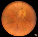
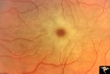
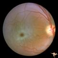
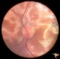
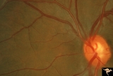
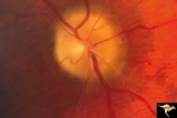
IF301 Post Giant Cell Arteritis Ischemic Papillopthy | Post giant cell arteritis ischemic papillopathy. Note shallow cupping without vascular displacement.1994. Also note the irregular focal arteriolar narrowing. Blind eye. Male. Anatomy: Optic disc. Pathology: Ischemic papillopathy from giant cell arteritis. Disease/ Diagnosis; Ischemic papillopathy fr... | Image |
| 532 |
 |
IIA101 Diffuse Atrophy | Primary or retrograde optic atrophy. It occurs from any injury to an optic nerve in the orbit or within the skull. Diffuse optic atrophy and blindness from nerve compression by pituitary tumor. Black woman. 1974. Note that retrograde optic atrophy can be associated with narrowed retinal arterioles i... | Image |
| 533 |
 |
IIA102a Diffuse Atrophy | Bilateral primary or retrograde optic atrophy from bilateral optic nerve sheath meningiomas. Pair with IIA1_2b. Right eye. 1984. Anatomy: Optic disc. Pathology: Bilateral optic nerve sheath meningiomas. Disease/ Diagnosis: Retrograde optic atrophy. Clinical: Bilateral visual loss. | Image |
| 534 |
 |
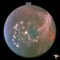
Ischemic Complication of Drusen | PP30a: right eye--buried drusen; PP30b: buried drusen with anterior ischemic optic neuropathy (AION) from complication of drusen of left eye. Ischemic complication of drusen in left eye. PP30c: 3 month follow-up: narrowed arterioles slightly pale disc with buried drusen. Anatomy: Optic disc. Patho... | Image |
| 535 |
 |
Ischemic Complication of Drusen | PP30a: right eye--buried drusen; PP3-b: buried drusen with anterior ischemic optic neuropathy (AION) from complication of drusen of left eye. Ischemic complication of drusen in left eye. PP30c: 3 month follow-up: narrowed arterioles slightly pale disc with buried drusen. Anatomy: Optic disc. Path... | Image |
| 536 |
 |
Ischemic Complication of Drusen | PP30a: right eye--buried drusen; PP30b: buried drusen with anterior ischemic optic neuropathy (AION) from complication of drusen of left eye. Ischemic complication of drusen in left eye. PP30c: 3 month follow-up: narrowed arterioles slightly pale disc with buried drusen. Anatomy: Optic disc. Patho... | Image |
| 537 |
 |
Late Complications of Drusen | PP33a: right disc shows pallor and small calcified crystals on the disc surface. PP33: left disc shows calcified specs on temporal sector of the disc. Florid drusen in young patients changes over time to assume this appearance. Anatomy: Optic disc. Pathology: Drusen of the optic disc. Disease/Dia... | Image |
| 538 |
 |
Late Complications of Drusen | PP33a: right disc shows pallor and small calcified crystals on the disc surface. PP33b: left disc shows calcified specs on temporal sector of the disc. Florid drusen in young patients changes over time to assume this appearance. Anatomy: Optic disc. Pathology: Drusen of the optic disc. Disease/Di... | Image |
| 539 |
 |
Macular Cherry Red Spots in Niemann-Pick disease | Close up view of macular cherry red spots in Niemann-Pick disease. Same patient as R2A2a. Anatomy: Retina. Pathology: Retinal ganglion cell accumulation of lipid. Disease/Diagnosis: Niemann-Pick disease. Clinical: Severe mental retardation and blindness. Fatal. | Image |
| 540 |
 |
Macular Cherry Red Spots in Niemann-Pick disease | Macular cherry red spots in Niemann-Pick disease. Same patient as R2A2b. Anatomy: Retina. Pathology: Retinal ganglion cell accumulation of lipid. Disease/Diagnosis: Niemann-Pick disease. Clinical: Severe mental retardation and blindness. Fatal. | Image |
| 541 |
 |
Macular Cherry Red Spots in Tay-Sachs disease | Macular cherry red spots in patient with Tay-Sachs disease. Anatomy: Retina. Pathology: Retinal ganglion cell accumulation of lipid. Disease/Diagnosis: Tay-Sachs disease. Clinical: Severe mental retardation and blindness. Fatal. | Image |
| 542 |
 |
Medullated Nerve Fibers with Papilledema | Left eye. papilledema only. Man with metastatic gastric carcinoma. Anatomy: Optic disc. Pathology: Papilledema. Disease/Diagnosis: Papilledema plus medullated nerve fibers. | Image |
| 543 |
 |
Medullated Nerve Fibers with Papilledema | Right eye. Papilledema superimposed upon medullated nerve fibers. Man with metastatic gastric carcinoma. Anatomy: Optic disc. Pathology: Papilledema. Disease/Diagnosis: Papilledema plus medullated nerve fibers. | Image |
| 544 |
 |
Multifocal Choroidopathy | Multifocal choroidopathy in a patient with uveitis. Anatomy: Retina. Disease/Diagnosis: Uveitis, Multifocal placoid pigment epitheliopathy. Clinical: Visual loss. | Image |
| 545 |
 |
Multifocal Choroidopathy | Multifocal choroidopathy in a patient with uveitis. Anatomy: Retina. Disease/Diagnosis: Uveitis, Multifocal placoid pigment epitheliopathy. Clinical: Visual loss. | Image |
| 546 |
 |
Multifocal Choroidopathy | Multifocal choroidopathy in a patient with uveitis. Anatomy: Retina. Disease/Diagnosis: Acute Multifocal Placoid Pigment Epitheliopathy (AMPPE). Clinical: Visual loss. | Image |
| 547 |
 |
Multifocal Choroidopathy | Multifocal choroidopathy in a patient with uveitis. Anatomy: Retina. Disease/Diagnosis: Acute Multifocal Placoid Pigment Epitheliopathy (AMPPE). Clinical: Visual loss. | Image |
| 548 |
 |
Multifocal Choroidopathy | Multifocal choroidopathy in a patient with uveitis. Anatomy: Retina. Disease/Diagnosis: Acute Multifocal Placoid Pigment Epitheliopathy (AMPPE). Clinical: Visual loss. | Image |
| 549 |
 |
Multifocal Choroidopathy | Multifocal choroidopathy in a patient with uveitis. Anatomy: Retina. Disease/Diagnosis: Acute Multifocal Placoid Pigment Epitheliopathy (AMPPE). Clinical: Visual loss. | Image |
| 550 |
 |
Multiple Sclerosis Slits and Thinning in Peripapillary (Retinal) Nerve Riber Layer | Multiple slit defect in the superior arcuate nerve fiber layer. Anatomy: Peripapillary nerve fiber layer. Pathology: Slit-like atrophy. Disease/Diagnosis: Multiple sclerosis optic neuropathy. Clinical: No symptoms. | Image |
| 551 |
 |
Multiple Sclerosis Slits and Thinning in Peripapillary (Retinal) Nerve Riber Layer | Multiple slit defect in the superior arcuate nerve fiber layer. Pair with IIB2_6b. Anatomy: Peripapillary nerve fiber layer. Pathology: Slit-like atrophy. Disease/Diagnosis: Multiple sclerosis optic neuropathy. Clinical: No symptoms. | Image |
| 552 |
 |
Multiple Sclerosis Slits and Thinning in Peripapillary (Retinal) Nerve Riber Layer | Multiple slit like defects in the inferior arcuate nerve fibers. Pair with IIB2_3b. Anatomy: Peripapillary nerve fiber layer. Pathology: Slit-like atrophy. Disease/Diagnosis: Multiple sclerosis optic neuropathy. Clinical: No symptoms. | Image |
| 553 |
 |
Multiple Sclerosis Slits and Thinning in Peripapillary (Retinal) Nerve Riber Layer | Multiple slit and wedge defects in the nerve fiber layer. Pair with IIB2_3a. Anatomy: Peripapillary nerve fiber layer. Pathology: Slit-like atrophy. Disease/Diagnosis: Multiple sclerosis optic neuropathy. Clinical: No symptoms. | Image |
| 554 |
 |
Multiple Sclerosis Slits and Thinning in Peripapillary (Retinal) Nerve Riber Layer | Multiple slit defect in the superior arcuate nerve fiber layer. Anatomy: Peripapillary nerve fiber layer. Pathology: Slit-like atrophy. Disease/Diagnosis: Multiple sclerosis optic neuropathy. Clinical: No symptoms. | Image |
| 555 |
 |
Multiple Sclerosis Slits and Thinning in Peripapillary (Retinal) Nerve Riber Layer | Multiple slit defect in the superior arcuate nerve fiber layer. Magnified. Pair with IIB2_6a. Anatomy: Peripapillary nerve fiber layer. Pathology: Slit-like atrophy. Disease/Diagnosis: Sclerosis optic neuropathy. Clinical: No symptoms. | Image |
| 556 |
 |
Multiple Sclerosis Slits and Thinning in Peripapillary (Retinal) Nerve Riber Layer | Left eye. Upper arcuate nerve fiber layer contains multiple low density slits. These indicate nerve fiber loss. Anatomy: Peripapillary nerve fiber layer. Pathology: Slit-like atrophy. Disease/Diagnosis: Multiple sclerosis optic neuropathy. Clinical: No symptoms. | Image |
| 557 |
 |
Multiple Sclerosis Slits and Thinning in Peripapillary (Retinal) Nerve Riber Layer | Multiple slit defect in the superior arcuate nerve fiber layer in a 13 year old boy. Right eye. Pair with IIB2_7a. Anatomy: Peripapillary nerve fiber layer. Pathology: Slit-like atrophy. Disease/Diagnosis: Multiple sclerosis optic neuropathy. Clinical: No symptoms. | Image |
| 558 |
 |
Multiple Sclerosis Slits and Thinning in Peripapillary (Retinal) Nerve Riber Layer | Multiple slit defect in the superior arcuate nerve fiber layer in a 13 year old boy. Pair with IIB2_7b. Anatomy: Peripapillary nerve fiber layer. Pathology: Slit-like atrophy. Disease/Diagnosis: Multiple sclerosis optic neuropathy. Clinical: No symptoms. | Image |
| 559 |
 |
Multiple Sclerosis Slits and Thinning in Peripapillary (Retinal) Nerve Riber Layer | Need magnification - Left eye - Peculiar punctate dotted surface of internal limiting membrane reflexes. Pairs with IIB2_01a & IIB2_02b. Anatomy: Peripapillary nerve fiber layer. Pathology: Slit-like atrophy. Disease/Diagnosis: Multiple sclerosis optic neuropathy. Clinical: No symptoms. | Image |
| 560 |
 |
Multiple Sclerosis Slits and Thinning in Peripapillary (Retinal) Nerve Riber Layer | Need magnification - Left eye - Inferior arcuate nerve fiber slits. Pairs with IIB2_01b & IIB2_01c. Anatomy: Peripapillary nerve fiber layer. Pathology: Slit-like atrophy. Disease/Diagnosis: Multiple sclerosis optic neuropathy. Clinical: No symptoms. | Image |
| 561 |
 |
Multiple Sclerosis Slits and Thinning in Peripapillary (Retinal) Nerve Riber Layer | Need magnification - Left eye - Inferior arcuate nerve fiber slits. Pairs with IIB2_01a & IIB2_01c. Anatomy: Peripapillary nerve fiber layer. Pathology: Slit-like atrophy. Disease/Diagnosis: Multiple sclerosis optic neuropathy. Clinical: No symptoms. | Image |
| 562 |
 |
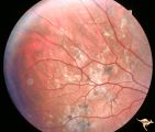
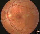
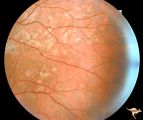
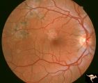
Neurofibromatosis-1 | Extensive retinal microvascular malformation involving both small and large retinal vessels. (Ref: BJO 2002:86, p282-284). Anatomy: Retina. Pathology: Retinal microvascular malformations. Disease/Diagnosis: Neurofibromatosis type 1. Clinical: No visual symptoms. | Image |
| 563 |
 |
Neurofibromatosis-1 | Normal appearing optic disc with dark pigmented choroidal nevi. The patient had NF-1 and had a subclinical optic glioma on the left eye. This is the right eye. Anatomy: Optic disc. Pathology: Choroidal nevus. Disease/Diagnosis: Neurofibromatosis type 1. Clinical: No visual symptoms. | Image |
| 564 |
 |
Neurofibromatosis-1 | Optic atrophy and hypoplasia of the optic disc associated with chiasmal glioma in a patient with NF-1. Anatomy: Optic disc. Pathology: Chiasmal glioma; Optic atrophy; Hypoplasia. Disease/Diagnosis: Neurofibromatosis type 1. Clinical: Proptosis; Blindness. | Image |
| 565 |
 |
Neurofibromatosis-1 | Fluorescein angiogram defines the extent of the microvascular malformation. Pair with R1_E5b. (Ref: BJO 2002:86, p282-284). Anatomy: Retina. Pathology: Retinal microvascular malformations. Disease/Diagnosis: Neurofibromatosis type 1. Clinical: No visual symptoms. Imaging: Fluorescein angiogram. | Image |
| 566 |
 |
Neurofibromatosis-1 | Retinal microvascular malformations in NF-1. Fundus picture shows a somewhat larger vertically running corkscrew malformation between two temporal retinal veins. Pair with R1_E5a. Anatomy: Retina. Pathology: Retinal microvascular malformations. Disease/Diagnosis: Neurofibromatosis type 1. Clinical: ... | Image |
| 567 |
 |
Neurofibromatosis-1 | Retinal microvascular malformations in NF-1 located between the disc and the macula. Anatomy: Retina. Pathology: Retinal microvascular malformations. Disease/Diagnosis: Neurofibromatosis type 1. Clinical: No visual symptoms. | Image |
| 568 |
 |
Neurofibromatosis-1 | Retinal microvascular malformations between optic disc and macula in NF-1. Anatomy: Retina. Pathology: Retinal microvascular malformations. Disease/Diagnosis: Neurofibromatosis type 1. Clinical: No visual symptoms. | Image |
| 569 |
 |
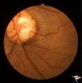
Neurofibromatosis-2 | CPERH (choroidal pigment epithelial retinal hamartoma) lesion in a patient with NF-2. Note the oblique superficial retinal traction folds running toward the center of the main lesion. 51 year old man. Anatomy: Retina. Pathology: Hamartoma. Disease/Diagnosis: Neurofibromatosis type 2. Clinical: Fiel... | Image |
| 570 |
 |
Neurofibromatosis-2 | Retinal tumor in NF-2 referred to as a CPERH (choroidal pigment epithelial retinal hamartoma). Patient, a 16 year old girl, had bilateral acoustic neurinomas. Pair with R1_F2b. Same eye. Anatomy: Optic disc; Retina. Pathology: Retinal hamartoma; Bilateral acoustic neurinoma. Disease/Diagnosis: Neuro... | Image |
| 571 |
 |
Neurofibromatosis-2 | Retinal tumor in NF-2 referred to as a CPERH (choroidal pigment epithelial retinal hamartoma). Patient, a 16 year old girl, had bilateral acoustic neurinomas. Pair with R1_F2a. Same eye. Anatomy: Optic disc; Retina. Pathology: Retinal hamartoma; Bilateral acoustic neurinoma. Disease/Diagnosis: Neuro... | Image |
| 572 |
 |
Neurofibromatosis-2 | This is the ocular fundus in a patient with NF-2 showing a preretinal membrane that extends from the temporal disc margin toward the macula. The optic disc shows low grade papilledema caused by one of the patient's acoustic neurinomas. The membrane has caused horizontal folds on the retinal surface.... | Image |
| 573 |
 |
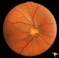
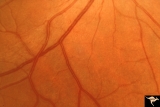
Normal Peripapillary Nerve Fiber Layer | Normal nerve fiber layer with Gunn's Dots visible in the upper arcuate fiber zone. This is a normal peripapillary nerve fiber layer in a young woman. Note the way the nerve fiber striations obscure and partially bury the small vessels running across them. Also note the interrupted surface reflex on ... | Image |
| 574 |
 |
Normal Peripapillary Nerve Fiber Layer | Example of 23 year old woman, healthy nerve fiber layer below her optic disc. This is a normal peripapillary nerve fiber layer in a young woman. Note the way the nerve fiber striations obscure and partially bury the small vessels running across them. Also note the interrupted surface reflex on arter... | Image |
| 575 |
 |
Normal Peripapillary Nerve Fiber Layer | Example of 23 year old woman, healthy nerve fiber layer below her optic disc. This is a normal peripapillary nerve fiber layer in a young woman. Note the way the nerve fiber striations obscure and partially bury the small vessels running across them. Also note the interrupted surface reflex on arter... | Image |
| 576 |
 |
Normal Peripapillary Nerve Fiber Layer | This is a normal peripapillary nerve fiber layer in a young woman. Note the way the nerve fiber striations obscure and partially bury the small vessels running across them. Also note the interrupted surface reflex on arteries due to interposed nerve fiber layer tissue. All these vessels are buried i... | Image |
| 577 |
 |
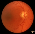
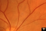
Normal Peripapillary Nerve Fiber Layer | This picture is a sample of a normal healthy nerve fiber layer temporal to the optic disc. Note how the nerve fiber layer obscures the small arteriolar branches running within it. Anatomy: Retina. Pathology: Normal healthy young retina. Disease/Diagnosis: Normal nerve fiber layer and retina. | Image |
| 578 |
 |
Normal Peripapillary Nerve Fiber Layer | Magnification of same disc shown in 1a. Whole fields of Gunn's Dots are visible. Note the way that the reflexes from the internal limiting membrane bend over vertically running arterioles. This is a tent-like effect. Gunn's Dots are footplates of Muller's cells and reflect light from the ophthalmosc... | Image |
| 579 |
 |
Ocular Hypertension | There is an argument about whether this is a pseudo nerve fiber layer defect, because defect does not reach the disc. The vessels transversing the defect are not exposed and there is no change in the visual field and no other slit like defects. Anatomy: Peripapillary nerve fiber layer. Pathology: Sl... | Image |
| 580 |
 |
Ocular Hypertension | Need color work to show superior slit 1972. Right eye. Ocular hypertension. No field defect recognized. Anatomy: Peripapillary nerve fiber layer. Pathology: Slit-like defects in the arcuate nerve fiber bundles. Disease/Diagnosis: Ocular hypertension. Clinical: Elevated intraocular pressure. | Image |
| 581 |
 |
Ocular Hypertension | 1972. Right eye. Ocular hypertension. No field defect recognized. Pair with IIB1b & c. Anatomy: Peripapillary nerve fiber layer. Pathology: Slit-like defects in the arcuate nerve fiber bundles. Disease/Diagnosis: Elevated intraocular pressure. Clinical: Elevated intraocular pressure. | Image |
| 582 |
 |
Ocular Hypertension | Multiple slit like defects in the upper arcuate nerve bundles. 1971. Anatomy: Peripapillary nerve fiber layer. Pathology: Slit-like defects in the arcuate nerve fiber bundles. Disease/Diagnosis: Elevated intraocular pressure. Clinical: Elevated intraocular pressure. | Image |
| 583 |
 |
Ocular Hypertension | Multiple slit defects in the upper arcuate bundles. 1972. Anatomy: Peripapillary nerve fiber layer. Pathology: Slit-like defects in the arcuate nerve fiber bundles. Disease/Diagnosis: Elevated intraocular pressure. Clinical: Elevated intraocular pressure. | Image |
| 584 |
 |
Ocular Hypertension | Chronic simple glaucoma. 1976. Magnified of IIB3_3a. Note slits in upper arcuate nerve fiber layer. Pair with IIB3_3a. Anatomy: Peripapillary nerve fiber layer. Pathology: Slit-like defects in the arcuate nerve fiber bundles. Disease/Diagnosis: Elevated intraocular pressure. Clinical: Elevated intra... | Image |
| 585 |
 |
Ocular Hypertension | Chronic simple glaucoma. 1976. Note slits in upper arcuate nerve fiber layer. Pair with IIB3_3b. Anatomy: Peripapillary nerve fiber layer. Pathology: Slit-like defects in the arcuate nerve fiber bundles. Disease/Diagnosis: Elevated intraocular pressure. Clinical: Elevated intraocular pressure. | Image |
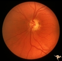
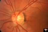
| 586 |
 |
P43a Asymmetrical Papilledema due to Brain Tumor | Right eye. Early papilledema. Incipient papilledema barely recognizable. Early papilledema due to posterior fossa meningioma in a boy. Anatomy: Optic disc. Pathology: Papilledema. Disease/ Diagnosis: Asymmetrical papilledema due to posterior fossa meningioma. | Image |
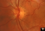
| 587 |
 |
P43b Asymmetrical Papilledema due to Brain Tumor | Left eye. Early papilledema. Clearly has papilledema. Early papilledema to posterior fossa meningioma in a boy. | Image |
| 588 |
 |
P50 Chronic Papilledema with Subretinal Neo-Vascular Network | Chronic papilledema with subretinal neo-vascular network. Pseudotumor. Anatomy: Optic disc. Pathology: Papilledema. Disease/ Diagnosis: Chronic papilledema with sub-retinal neovascular network. | Image |
| 589 |
 |
P52a Asymmetric Papilledema with Choroidal Folds | Left eye. Choroidal folds with no papilledema. Asymmetric papilledema with choroidal folds. Bilateral choroidal folds from elevated intracranial pressure. 52a. Anatomy: Optic disc. Pathology: Papilledema. Disease/ Diagnosis: Asymmetric - No papilledema with choroidal folds | Image |
| 590 |
 |
P52b Asymmetric Papilledema with Choroidal Folds | Right eye shows papilledema. Asymmetric papilledema with choroidal folds. Bilateral choroidal folds from elevated intracranial pressure. Anatomy: Optic disc. Pathology: Papilledema. Disease/ Diagnosis: Chronic papilledema with choroidal folds. | Image |
| 591 |
 |
P55a Chronic Papilledema, Late Stage | Right eye. Optic atrophy secondary to chronic papilledema. Glioblastoma. Chemotherapy patient. Anatomy: Optic disc. Pathology: Papilledema. Disease/ Diagnosis: Chronic papilledema with atrophy. | Image |
| 592 |
 |
P55b Chronic Papilledema, Late Stage | Left eye. Optic atrophy secondary to chronic papilledema, late stage with retinal choroidal bypass vein. Opticocilliary shunt. Glioblastoma, chemotherapy patient. Anatomy: Optic disc. Pathology: Papilledema. Disease/ Diagnosis: Chronic papilledema with atrophy. | Image |
| 593 |
 |
Papilledema due to Brain Tumor - Natural History | Right eye 33 months after presentation. Atrophy appears about the same. Notice especially the narrowing of retinal arterioles. Visual loss is severe in both eyes. Papilledema due to brain tumor - 3 year natural history. Patient refused treatment. Man. Anatomy: Optic disc. Pathology: Papilledema. Dis... | Image |
| 594 |
 |
Papilledema due to Brain Tumor - Natural History | Right eye at 27 months after presentation. Atrophy is more profound in both eyes. Papilledema due to brain tumor - 3 year natural history. Patient refused treatment. Man. Anatomy: Optic disc. Pathology: Papilledema. Disease/Diagnosis: Papilledema from acoustic neuronoma. | Image |
| 595 |
 |
Papilledema due to Brain Tumor - Natural History | Left eye at 25 months after presentation. Atrophy is more profound in both eyes. Papilledema due to brain tumor - 3 year natural history. Patient refused treatment. Man. Anatomy: Optic disc. Pathology: Papilledema. Disease/Diagnosis: Papilledema from acoustic neuronoma. | Image |
| 596 |
 |
Papilledema due to Brain Tumor - Natural History | Right eye 23 months after presentation. Atrophy is replacing papilledema in both eyes. Papilledema due to brain tumor - 3 year natural history. Patient refused treatment. Man. Anatomy: Optic disc. Pathology: Papilledema. Disease/Diagnosis: Papilledema from acoustic neuronoma. | Image |
| 597 |
 |
Papilledema due to Brain Tumor - Natural History | Left eye 23 months after presentation. Atrophy is replacing papilledema in both eyes. Papilledema due to brain tumor - 3 year natural history. Patient refused treatment. Man. Anatomy: Optic disc. Pathology: Papilledema. Disease/Diagnosis: Papilledema from acoustic neuronoma. | Image |
| 598 |
 |
Papilledema due to Brain Tumor - Natural History | Left eye. 3.5 years after presentation. Atrophy appears about the same. Note especially the narowing of retinal arterioles. Visual loss is profound in both eyes. Note the horizontal retinal folds. Papilledema due to brain tumor - 3 year natural history. Patient refused treatment. Man. | Image |
| 599 |
 |
Papilledema due to Brain Tumor - Natural History | Right eye at presentation. Papilledema due to acoustic neuronoma(tumor)- 3 year natural history. Patient refused treatment. Man. Anatomy: Optic disc. Pathology: Papilledema. Disease/Diagnosis: Papilledema from acoustic neuronoma. | Image |
| 600 |
 |
Papilledema due to Brain Tumor - Natural History | Left eye at presentation. Papilledema due to acoustic neuronoma(tumor) - 3 year natural history. Patient refused treatment. Man. Anatomy: Optic disc. Pathology: Papilledema. Disease/Diagnosis: Papilledema from acoustic neuronoma. | Image |