Best known for his world-renowned neuro-ophthalmology unit based at the University of California, San Francisco, William Hoyt, MD collected here more than 850 of his best images covering a wide range of disorders.
William F. Hoyt, MD, Professor Emeritus of Ophthalmology, Neurology and Neurosurgery, Department of Ophthalmology, University of California, San Francisco.
NOVEL: https://novel.utah.edu/
TO
Filters: Collection: "ehsl_novel_wfh"
| Title | Description | Type | ||
|---|---|---|---|---|
| 126 |
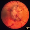 |
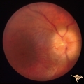
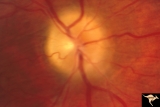
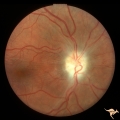
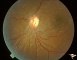
Bilateral Papilledema from Occipital Tumor | Right eye. Bilateral hemorrhagic papilledema. Occipital glioma. Right hemianopia. Woman. Anatomy: Optic disc. Pathology: Papilledema. Disease/Diagnosis: Hemorrhagic papilledema from occipital glioma. | Image |
| 127 |
 |
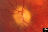
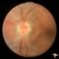
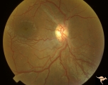
Papilledema due to Brain Tumor - Natural History | Right eye 33 months after presentation. Atrophy appears about the same. Notice especially the narrowing of retinal arterioles. Visual loss is severe in both eyes. Papilledema due to brain tumor - 3 year natural history. Patient refused treatment. Man. Anatomy: Optic disc. Pathology: Papilledema. Dis... | Image |
| 128 |
 |
Papilledema due to Brain Tumor - Natural History | Right eye at 27 months after presentation. Atrophy is more profound in both eyes. Papilledema due to brain tumor - 3 year natural history. Patient refused treatment. Man. Anatomy: Optic disc. Pathology: Papilledema. Disease/Diagnosis: Papilledema from acoustic neuronoma. | Image |
| 129 |
 |
Papilledema due to Brain Tumor - Natural History | Left eye at 25 months after presentation. Atrophy is more profound in both eyes. Papilledema due to brain tumor - 3 year natural history. Patient refused treatment. Man. Anatomy: Optic disc. Pathology: Papilledema. Disease/Diagnosis: Papilledema from acoustic neuronoma. | Image |
| 130 |
 |
Papilledema due to Brain Tumor - Natural History | Right eye 23 months after presentation. Atrophy is replacing papilledema in both eyes. Papilledema due to brain tumor - 3 year natural history. Patient refused treatment. Man. Anatomy: Optic disc. Pathology: Papilledema. Disease/Diagnosis: Papilledema from acoustic neuronoma. | Image |
| 131 |
 |
Papilledema due to Brain Tumor - Natural History | Left eye 23 months after presentation. Atrophy is replacing papilledema in both eyes. Papilledema due to brain tumor - 3 year natural history. Patient refused treatment. Man. Anatomy: Optic disc. Pathology: Papilledema. Disease/Diagnosis: Papilledema from acoustic neuronoma. | Image |
| 132 |
 |
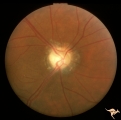
Papilledema due to Brain Tumor - Natural History | Right eye at presentation. Papilledema due to acoustic neuronoma(tumor)- 3 year natural history. Patient refused treatment. Man. Anatomy: Optic disc. Pathology: Papilledema. Disease/Diagnosis: Papilledema from acoustic neuronoma. | Image |
| 133 |
 |
Papilledema due to Brain Tumor - Natural History | Left eye at presentation. Papilledema due to acoustic neuronoma(tumor) - 3 year natural history. Patient refused treatment. Man. Anatomy: Optic disc. Pathology: Papilledema. Disease/Diagnosis: Papilledema from acoustic neuronoma. | Image |
| 134 |
 |
Papilledema due to Brain Tumor - Natural History | Right eye 3.5 years after presentation. Atrophy appears about the same. Note especially the narrowing of retinal arterioles. Visual loss is prfound in both eyes. Note the horizontal retinal folds. Papilledema due to brain tumor - 3 year natural history. Patient refused treatment. Man. | Image |
| 135 |
 |
Papilledema due to Brain Tumor - Natural History | Left eye at 33 months after presentation. Atrophy appears about the same. Note especially the narrowing of retinal arterioles. Visual loss is severe in both eyes. Papilledema due to brain tumor - 3 year natural history. Patient refused treatment. Man. | Image |
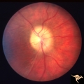
| 136 |
 |
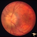
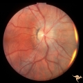
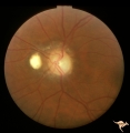
Bilateral Papilledema with Pseudotumor Cerebri | Right eye. Mild bilateral papilledema in a 7 year old boy. Cause of swelling unknown. Growth failure treated with thyroid medication. Anatomy: Optic disc. Pathology: Bilateral papilledema. Disease/Diagnosis: Intracranial hypertension due to treatment of growth failure with thyroid medicaltion. Clini... | Image |
| 137 |
 |
Bilateral Papilledema with Pseudotumor Cerebri | Left eye. Mild bilateral papilledema in a 7 year old boy. Cause of swelling unknown. Growth failure treated with thyroid medication. Anatomy: Optic disc. Pathology: Bilateral papilledema. Disease/Diagnosis: Intracranial hypertension due to treatment of growth failure with thyroid medication. Clinica... | Image |
| 138 |
 |
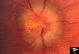
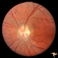
Bilateral Papilledema with Pseudotumor Cerebri | Chronic appearance of swelling in right eye. 29 year old woman. Bilateral papilledema. Anatomy: Optic disc. Pathology: Bilateral papilledema. Disease/Diagnosis: Intracranial hypertension due to treatment of growth failure with thyroid medication. Clinical: symptoms: headache, signs: bilateral papill... | Image |
| 139 |
 |
ID01 Post Papilledema Gliosis | Post papilledema milky gliosis with arteriolar constriction, 1982, right eye, pair with ID_2. Anatomy: Optic disc. Pathology: Post papilledema atrophy and gliosis due to huge anterior communicating artery aneurysm. Disease/ Diagnosis: Elevated intracranial pressure from aneurysm. Clinical: Diminishe... | Image |
| 140 |
 |
ID02 Post Papilledema Gliosis | Post papilledema milky gliosis with arteriolar constriction and atrophy, 1982, left eye, pair with ID_1. Anatomy: Optic disc. Pathology: Post papilledema atrophy and gliosis due to huge anterior communicating artery aneurysm. Disease/ Diagnosis: Elevated intracranial pressure from aneurysm. Clinical... | Image |
| 141 |
 |
ID03a Post Papilledema Atrophy with Marked Gliosis | Post papilledema atrophy with marked gliosis in a patient with pseudotumor cerebri. Patient weighed over 300 pounds. Right eye blind. 1981. Right eye. Pair with ID_3b. Anatomy: Optic disc. Pathology: Post papilledema atrophy and gliosis from long standing elevated intracranial pressure. Disease/ Dia... | Image |
| 142 |
 |
ID03b Post Papilledema Atrophy with Marked Gliosis | Post papilledema atrophy with marked gliosis in a patient with pseudotumor cerebri. Patient weighed over 300 pounds. Left eye has visual field defects. 1981, right eye, pair with ID_3a. Anatomy: Optic disc. Pathology: Post papilledema atrophy and gliosis from long standing elevated intracranial pres... | Image |
| 143 |
 |
ID04a Post Papilledema Atrophy with Marked Gliosis | Post papilledema atrophy with marked gliosis in a patient with pseudotumor cerebri, 1985, right eye, pair with ID_4b, Note "high water" marks in peripapillary pigment epithelial layer. Anatomy: Optic disc. Pathology: Post papilledema atrophy and gliosis from long standing elevated intracranial press... | Image |
| 144 |
 |
ID04b Post Papilledema Atrophy with Marked Gliosis | Post papilledema atrophy with marked gliosis in a patient with pseudotumor. Nasal ovoid absence of the retinal pigment epithelium. Presumably a defect from the long standing papilledema. 1985,. Right eye, pair with ID_4a. Anatomy: Optic disc. Pathology: Post papilledema atrophy and gliosis from long... | Image |
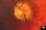
| 145 |
 |
ID05a Post Papilledema Optic Atrophy from Pseudotumor Cerebri | Left eye, October 1999, Post papilledema optic atrophy from pseudotumor cerebri. Note optociliary veins in both discs. Gliosis and partial pallor following long standing papilledema and intracranial pressure. Anatomy: Optic disc. Pathology: Post papilledema atrophy and gliosis from long standing el... | Image |
| 146 |
 |
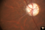
ID05b Post Papilledema Optic Atrophy from Pseudotumor Cerebri | Right eye, October 1999, Post papilledema optic atrophy from pseudotumor cerebri. Note optociliary veins in both discs. Gliosis and partial pallor following long standing papilledema and intracranial pressure. Anatomy: Optic disc. Pathology: Post papilledema atrophy and gliosis from long standing el... | Image |
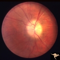
| 147 |
 |
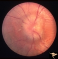
ID06 Post Papilledema Optic Atrophy with Arteriolar Sheathing and Optociliary Veins | 1989. Post papilledema optic atrophy with arteriolar sheathing and optociliary veins. Anatomy: Optic disc. Pathology: Long standing effects of intracranial pressure. Clinical: Blindness. | Image |
| 148 |
 |
ID07 Post Papilledema Optic Atrophy | Post papilledema optic atrophy with gliosis and arteriolar narrowing. 1994. Anatomy: Optic disc. Pathology: Residue of long standing papilledema. Clinical: Visual loss. | Image |