Best known for his world-renowned neuro-ophthalmology unit based at the University of California, San Francisco, William Hoyt, MD collected here more than 850 of his best images covering a wide range of disorders.
William F. Hoyt, MD, Professor Emeritus of Ophthalmology, Neurology and Neurosurgery, Department of Ophthalmology, University of California, San Francisco.
NOVEL: https://novel.utah.edu/
TO
| Title | Description | Type | ||
|---|---|---|---|---|
| 101 |
 |
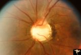
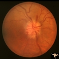
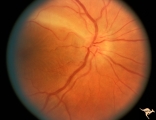
Buried Drusen with Choroidal Retinal Scar | Right eye: Buried drusen; probable complication of peripapillary hemorrhage at 7:00. Anatomy: Optic disc. Pathology: Drusen of the optic disc. Disease/Diagnosis: Drusen of the optic disc. Clinical notes: Enlarged blind spot. | Image |
| 102 |
 |
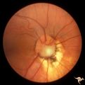
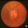
Buried Drusen with Sub-retinal Neovascular Net | Buried drusen with sub-retinal neovascular net. Both PP29a and PP29b are left eye: 17 year old girl; Visual acuity 10/400. Anatomy: Optic disc. Pathology: Drusen of the optic disc. Disease/Diagnosis: Drusen of the optic disc. Clinical notes: Loss of central vision due to subretinal neovascularizatio... | Image |
| 103 |
 |
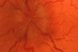
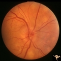
Buried Drusen with Sub-retinal Neovascular Net | Buried drusen with sub-retinal neovascular net. This is the same left eye. Appearance of the central retina of the left eye. Both PP29a & b are left eye: 17 year old girl; Visual acuity 10/400. Anatomy: Optic disc. Pathology: Drusen of the optic disc. DIsease/Diagnosis: Drusen of the optic disc. Cl... | Image |
| 104 |
 |
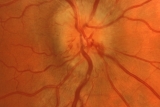
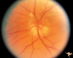
C01 Pits of the Optic Disc | Right eye. Very large inferior temporal optic pit. Congenital. Woman. Anatomy: Optic disc. | Image |
| 105 |
 |
C03 Pits of the Optic Disc | Central optic pit. Left eye. Anatomy: Optic disc. | Image |
| 106 |
 |
C04 Pits of the Optic Disc | Right eye. Man. Large temporal pit. Macular detachment. Anatomy: Optic disc. | Image |
| 107 |
 |
C05 Pits of the Optic Disc | Right eye. Pigmented pit. Woman. Anatomy: Optic disc. | Image |
| 108 |
 |
C06 Pits of the Optic Disc | Right eye. Temporal pit. 6 year old with see-saw nystagmus. Anatomy: Optic disc. Clinical: Six-year old with see-saw nystagmus. | Image |
| 109 |
 |
C07 Pits of the Optic Disc | Left eye. Temporal pit. Man. Anatomy: Optic disc. | Image |
| 110 |
 |
C08 Pits of the Optic Disc | Left eye. Large cavitary anomaly (pit). Man. 20/100 visual acuity. Superior nasal visual field defect. May not have a central retinal artery. Anatomy: Optic disc. Clinical: Man. 20/100 visual acuity. Superior nasal visual field defect. | Image |
| 111 |
 |
C09 Pits of the Optic Disc | Pit with peripapillary choroidal defect. Right eye. Dwarfed boy. May not have a central retinal artery. Same patient as C_10. Anatomy: Optic disc. | Image |
| 112 |
 |
C10 Pits of the Optic Disc | Disc malformation. Abortive cavitary anomaly. Left eye. Dwarfed boy. Same patient as C_9. Anatomy: Optic disc. | Image |
| 113 |
 |
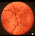
C101 Papillitis, Retrobulbar Neuritis | Resolved. Associated polycythemia. Papillitis after flu in patient with polycythemia. Homosexual male. Anatomy: Optic disc. Pathology: Axoplasmic stasis due to inflammation. Disease/ Diagnosis: Post infectious papillitis. Clinical: Visual loss after the flu.. | Image |
| 114 |
 |
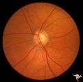
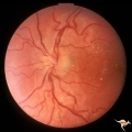
C102 Papillitis, Retrobulbar Neuritis | Inflammatory papillitis in 25 year old woman. Resolved completely. Anatomy: Optic disc. Pathology: Axoplasmic stasis due to inflammation. Disease/ Diagnosis: Inflammatory optic papillitis. Clinical: Visual loss. | Image |
| 115 |
 |
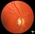
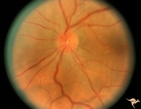
C103 Papillitis, Retrobulbar Neuritis | Optic neuritis in infectious mononucleosis. Anatomy: Optic disc. Pathology: Axoplasmic stasis due to inflammation. Disease/ Diagnosis: Optic neuritis with mononucleosis or Epstein Barr Virus. Clinical: Visual loss associated with mononucleosis. | Image |
| 116 |
 |
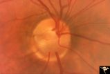
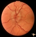
C104 Papillitis, Retrobulbar Neuritis | Post infectious papillitis with macular exudate. Anatomy: Optic disc macula. Pathology: Axoplasmic stasis due to inflammation with lipid deposit in Henle's layer. Disease/ Diagnosis: Post infectious papillitis/optic neuritis. Clinical: Visual loss after infection. | Image |
| 117 |
 |
C105 Disc Edema with Systemic Lupus | Mild disc edema blurs the inferior disc margin. Flourescein angiogram in D1_06. Same patient as D1_06 an D1_07. Man. Anatomy: Optic disc. Pathology: Axoplasmic stasis due to vasculitis (Lupus). Disease/ Diagnosis: Lupus papillopathy. | Image |
| 118 |
 |
C106 Papillitis, Retrobulbar Neuritis | Papillitis with recovery of vision. Woman acupuncturist. Anatomy: Optic disc. Pathology: Axoplasmic stasis after inflammation. Disease/ Diagnosis: Optic neuritis/optic papillitis. Clinical: Visual loss with recovery. | Image |
| 119 |
 |
C107 Papillitis, Retrobulbar Neuritis | Man with bilateral papillitis. Right eye. Pair with C1_08. Cause unknown. Visual field showed central scotomas. Anatomy: Optic disc. Pathology: Axoplasmic stasis due to inflammation. Disease/ Diagnosis: Neuritis of the optic nerve. Clinical: Visual loss. | Image |
| 120 |
 |
C108 Papillitis, Retrobulbar Neuritis | Man with bilateral papillitis. Left eye. Pair with C1_07. Cause unknown. Visual field shows central scotoma. Anatomy: Optic disc. Pathology: Axoplasmic stasis due to inflammation. Disease/ Diagnosis: Optic neuritis / Optic papillitis. Clinical: Visual loss. | Image |
| 121 |
 |
C109 Papillitis, Retrobulbar Neuritis | Optic papillitis after wasp sting. 57 year old woman. Right eye. Anatomy: Optic disc. Pathology: Axoplasmic stasis due to inflammation. Disease/ Diagnosis: Optic neuritis after wasp sting. Clinical: Visual loss after wasp sting. | Image |
| 122 |
 |
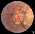
C110 Papillitis, Retrobulbar Neuritis | AIDs papillitis. Segmental. Note inflammatory focus on temporal side of disc. 29 year old homosexual male. Visual field shows huge blind spot. Anatomy: Optic disc. Pathology: Axoplasmic stasis due to inflammation. Disease/ Diagnosis: AIDS papillitis / AIDS Optic neuritis. Clinical: Visual symptoms d... | Image |
| 123 |
 |
C111 Papillitis, Retrobulbar Neuritis | AIDS papillitis. Male. Anatomy: Optic disc. Pathology: Axoplasmic stasis due to inflammation. Disease/ Diagnosis: AIDS papillitis. Clinical: Visual symptoms. | Image |
| 124 |
 |
C112 Papillitis, Retrobulbar Neuritis | Woman with herpes. Acute retinal necrosis with papillitis and arcuate neuro-retinitis. Right eye. Pair with C1_13. Reference: Margolis T, Irvine AR, Hoyt WF, Hyman R. Acute retinal necrosis syndrome presenting with papillitis and arcuate neuroretinitis. Ophthalmology. 1988 Jul;95(7):937-40. Anatomy:... | Image |
| 125 |
 |
C113 Papillitis, Retrobulbar Neuritis | Woman with herpes. Acute retinal necrosis with papillitis an arcuate neuro-retinitis. Right eye. Notice the large arcuate defect extending fromt he disc to the retina of retinal necrosis. Pair with C1_12. Anatomy: Optic disc; Retina. Pathology: Axoplasmic stasis due to inflammation; Retinal necrosis... | Image |