Best known for his world-renowned neuro-ophthalmology unit based at the University of California, San Francisco, William Hoyt, MD collected here more than 850 of his best images covering a wide range of disorders.
William F. Hoyt, MD, Professor Emeritus of Ophthalmology, Neurology and Neurosurgery, Department of Ophthalmology, University of California, San Francisco.
NOVEL: https://novel.utah.edu/
TO
Filters: Collection: "ehsl_novel_wfh"
| Title | Description | Type | ||
|---|---|---|---|---|
| 401 |
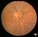 |
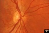
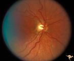
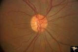
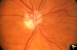
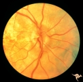
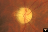
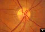
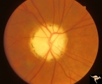
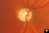
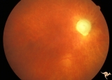
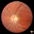
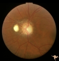
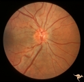
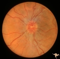
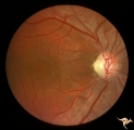
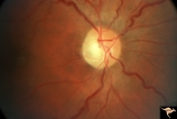
H77 Inferior Segmental Optic Hypoplasia (ISOH) | ISOH. Superior visual field defect. Inferior choroidal crescent. Anatomy: Optic disc. Pathology: Inferior segmental optic hypoplasia (ISOH). Disease/ Diagnosis: Congenital anomaly. | Image |
| 402 |
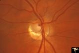 |
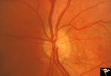
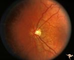
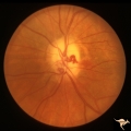
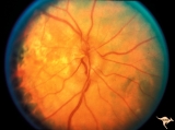
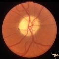
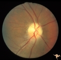
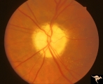
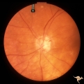
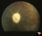
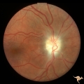
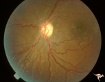
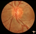
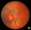
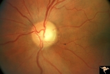
H78 Inferior Segmental Optic Hypoplasia (ISOH) | ISOH with inferior choroidal crescent. Patient had superior visual field defect. Anatomy: Optic disc. Pathology: Inferior segmental optic hypoplasia (ISOH). Disease/ Diagnosis: Congenital anomaly. | Image |
| 403 |
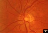 |
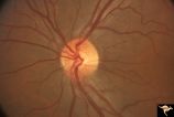
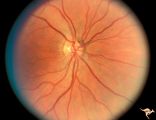
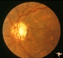
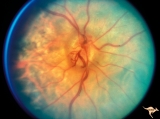
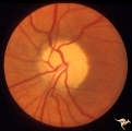
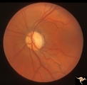
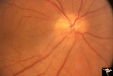
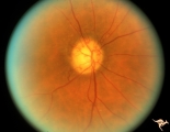
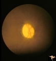
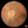
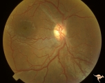
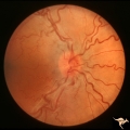
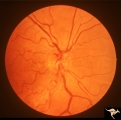
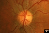
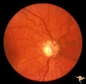
H79 Inferior Segmental Optic Hypoplasia (ISOH) | ISOH. Anatomy: Optic disc. Pathology: Inferior segmental optic hypoplasia (ISOH). Disease/ Diagnosis: Congenital anomaly. | Image |
| 404 |
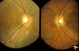 |
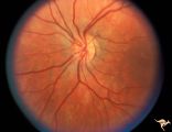
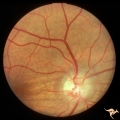
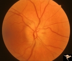
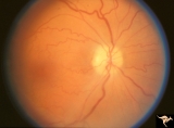
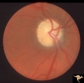
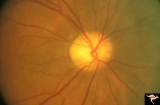
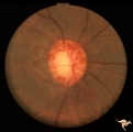
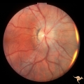
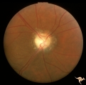
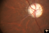
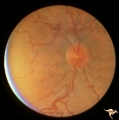
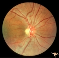
H80 Chiasmal Hemioptic Hypoplasia | Discs show striking nasal hypoplasia and band atrophy. DeMorsier synrome. Congenital bitemporal hemianopia with see-saw nystagmus. Note vertically oral shape of these hypoplastic nerves. The CT scan showed the median bar of the chiasm in this patient is totally hypoplastic. Anatomy: Optic disc. Path... | Image |
| 405 |
 |
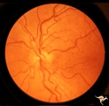
H81 Chiasmal Hemioptic Hypoplasia | De Morsier synrome with congenital bitemporal hemianopia. Right eye. Note nasal hypoplasia of the right optic disc. Same patient as H_82. Anatomy: Optic disc. Pathology: Chiasmal hemioptic hypoplasia. Disease/ Diagnosis: Congenital anomaly involving chiasm | Image |
| 406 |
 |
H82 Chiasmal Hemioptic Hypoplasia | De Morsier synrome with congenital bitemporal hemianopia. Left eye. Same patient as H_81. Anatomy: Optic disc. Pathology: Chiasmal hemioptic hypoplasia. Disease/ Diagnosis: Congenital anomaly involving chiasm. | Image |
| 407 |
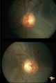 |
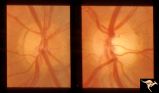
H83 Chiasmal Hemioptic Hypoplasia | De Morsier synrome with congenital bitemporal hemianopia. Note nasal hypoplasia of both optic discs. Left eye above, right eye below. Anatomy: Optic disc. Pathology: Chiasmal hemioptic hypoplasia. Disease/ Diagnosis: Congenital anomaly involving chiasm. | Image |
| 408 |
 |
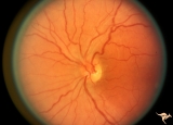
H84 Chiasmal Hemioptic Hypoplasia | Congenital bitemporal hemianopia with marked bi-nasal hypoplasia. Left eye. 17 year old male. Same patient as H_85. Anatomy: Optic disc. Pathology: Chiasmal hemioptic hypoplasia. Disease/ Diagnosis: Congenital anomaly involving chiasm. | Image |
| 409 |
 |
H85 Chiasmal Hemioptic Hypoplasia | Congenital bitemporal hemianopia with marked bi-nasal hypoplasia. Right eye. 17 year old male. Same patient as H_84. Anatomy: Optic disc. Pathology: Chiasmal hemioptic hypoplasia. Disease/ Diagnosis: Congenital anomaly involving chiasm. | Image |
| 410 |
 |
H86 Chiasmal Hemioptic Hypoplasia | Congenital bitemporal hemianopia with nasal hypoplasia. 24 year old man. Same patient as H_87. Anatomy: Optic disc. Pathology: Chiasmal hemioptic hypoplasia. Disease/ Diagnosis: Congenital anomaly involving chiasm. | Image |
| 411 |
 |
H87 Chiasmal Hemioptic Hypoplasia | Congenital bitemporal hemianopia with nasal hypoplasia. 24 year old man. Same patient as H_86. Anatomy: Optic disc. Pathology: Chiasmal hemioptic hypoplasia. Disease/ Diagnosis: Congenital anomaly involving chiasm. | Image |
| 412 |
 |
H88 Chiasmal Hemioptic Hypoplasia | Nasal hypoplasia with temporal hemianopia from a congenital Rathke Pouch Cyst. Anatomy: Optic disc. Pathology: Chiasmal hemioptic hypoplasia. Disease/ Diagnosis: Congenital anomaly involving chiasm. | Image |
| 413 |
 |
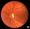
H89 Occipital Hemianoptic Hypoplasia | Diagram of homonymous hemioptic hypoplasia showing pattern of preserved nerve fibers. Homonymous hemioptic hypoplasia. Fundoscopic features in standard and red-free illumination in three patients with congenital hemiplegia. Anatomy: Optic disc. Pathology: Occipital hemianoptic hypoplasia. Disease/ D... | Image |
| 414 |
 |
H90 Occipital Hemianoptic Hypoplasia | Note left disc (right side of image) is the eye with temporal field defect. Shows band atrophy. Anatomy: Optic disc. Pathology: Occipital hemianoptic hypoplasia. Congenital defect of the occipital lobe. | Image |
| 415 |
 |
H91 Occipital Hemianoptic Hypoplasia | Left eye with temporal field defect shows trans-synaptic band atrophy. Same patient as H_92. Anatomy: Optic disc. Pathology: Occipital hemianoptic hypoplasia. Disease/ Diagnosis: Congenital defect of the occipital lobe. | Image |
| 416 |
 |
H92 Occipital Hemianoptic Hypoplasia | Right eye. Same patient as H_91. Anatomy: Optic disc. Pathology: Occipital hemianoptic hypoplasia. Disease/ Diagnosis: Congenital defect of the occipital lobe. | Image |
| 417 |
 |
H93 Occipital Hemianoptic Hypoplasia | Visual field. Left eye. Right inferior homonymous. Same patient as H_94, H_95, H_96, H_97. Anatomy: Optic disc. Pathology: Occipital hemianoptic hypoplasia. Disease/ Diagnosis: Congenital defect of the occipital lobe. Imaging: MRI scan - See slide H97. | Image |
| 418 |
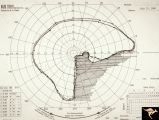 |
H94 Occipital Hemianoptic Hypoplasia | Visual field. Right eye. Quatrantanopia. Same patient as H_93, H_95, H_96, H_97. Anatomy: Optic disc. Pathology: Occipital hemianoptic hypoplasia. Disease/ Diagnosis: Congenital defect of the occipital lobe. Imaging: MRI scan - See slide H97. | Image |
| 419 |
 |
H95 Occipital Hemianoptic Hypoplasia | Right eye with temporal field defect shows trans-synaptic band atrophy. Absence of nasal nerve fibers. Same patient as H_93, H_94, H_96, H_97. Anatomy: Optic disc. Pathology: Occipital hemianoptic hypoplasia. Disease/ Diagnosis: Congenital defect of the occipital lobe. Imaging: MRI scan - See slide ... | Image |
| 420 |
 |
H96 Occipital Hemianoptic Hypoplasia | Left eye has nasal quadrantic field defect. Same patient as H_93, H_94, H_95, H_97. Anatomy: Optic disc. Pathology: Occipital hemianoptic hypoplasia. Disease/ Diagnosis: Congenital defect of the occipital lobe. Imaging: MRI scan - See slide H97. | Image |
| 421 |
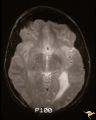 |
H97 Occipital Hemianoptic Hypoplasia | MRI scan shows left occipital lobe periventricular leuko-melacia. Same patient as H_93, H_94, H_95, H_96. Anatomy: Optic disc. Pathology: Occipital hemianoptic hypoplasia. DIsease/ Diagnosis: Congenital defect of the occipital lobe. Imaging: MRI scan. | Image |
| 422 |
 |
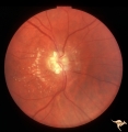
Hemorrhagic Complication of Drusen | PP31a, left and PP31, right taken in April. PP31c: left taken after an interval of 2 months. Hemorrhage. Hemorrhagic complications of drusen. 15 year old boy. Anatomy: Optic disc. Pathology: Drusen of the optic disc. Disease/Diagnosis: Drusen of the optic disc. Clinical: Patient complained of blurre... | Image |
| 423 |
 |
Hemorrhagic Complication of Drusen | PP31a, left and PP31, right taken in April. PP31c: left taken after an interval of 2 months. Hemorrhage. Hemorrhagic complications of drusen. 15 year old boy. Anatomy: Optic disc. Pathology: Drusen of the optic disc. Disease/Diagnosis: Drusen of the optic disc. Clinical: Hemorrhage in drusen disc. | Image |
| 424 |
 |
Hemorrhagic Complication of Drusen | PP31a, left and PP31, right taken in April. PP31c: left taken after an interval of 2 months. Hemorrhage. Hemorrhagic complications of drusen. 15 year old boy. Anatomy: Optic disc. Pathology: Drusen of the optic disc. Disease/Diagnosis: Drusen of the optic disc. Clinical: Drusen. | Image |
| 425 |
 |
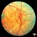
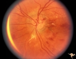
Hereditary Macular Degenerative Disease with Spastic Paraplegia | Hereditary macular degenerative disease with Patient has spastic paraplegia associated with hereditary macular degenerative disease. Anatomy: Retina. Pathology: Cerebellar spinal degenerative disease. Disease/Diagnosis: Retinitis pigmentosa with spinal degeneration. Clinical: Hereditary spastic para... | Image |
| 426 |
 |
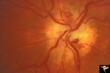
IA01 Atrophy with Optociliary Veins | 1994, perioptic nerve sheath meningioma, right eye, Optociliary vein dumping into disc edge at 4:00. Anatomy: Optic disc. Pathology: Optociliary vein. Disease/ Diagnosis: Perioptic nerve sheath meningioma. Clinical: Progressive visual loss | Image |
| 427 |
 |
IA02 Atrophy with Optociliary Veins | 1971, left eye, perioptic nerve sheath meningioma, notice how vein dumps into adjacent choroid at 3:00. The darker venous blood can be seen at the disc edge. Anatomy: Optic disc. Pathology: Optociliary vein. Disease/ Diagnosis: Perioptic nerve sheath meningioma. Clinical: Progressive visual loss. | Image |
| 428 |
 |
IA03 Atrophy with Optociliary Veins | 1974, left eye, perioptic nerve sheath meningioma, blind eye. Optociliary bypass veins in the nasal disc tissue. Anatomy: Optic disc. Pathology: Optociliary vein. DIsease/ Diagnosis: Perioptic nerve sheath meningioma. Clinical: Blind eye. | Image |
| 429 |
 |
IA04 Atrophy with Optociliary Veins | 1981, right eye, perioptic nerve sheath meningioma with optociliary bypass vein. Notice horizontal choroidal folds in the retina from posterior tumor pressure. Anatomy: Optic disc. Pathology: Optociliary vein. Disease/ Diagnosis: Perioptic nerve sheath meningioma. Clinical: Blind eye. | Image |
| 430 |
 |
IA05 Atrophy with Optociliary Veins | 1971, right eye, perioptic nerve sheath meningioma with optociliary bypass veins on the upper half of the disc. Anatomy: Optic disc. Pathology: Optociliary vein. Disease/ Diagnosis: Perioptic nerve sheath meningioma. Clinical: Blind eye. | Image |
| 431 |
 |
IA06 Atrophy with Optociliary Veins | 1979, left eye, perioptic nerve sheath meningioma with optociliary bypass veins. Anatomy: Optic disc. Pathology: Optociliary vein. Disease/ Diagnosis: Perioptic nerve sheath meningioma. Clinical: Blind eye. | Image |
| 432 |
 |
IA07 Atrophy with Optociliary Veins | Left eye, perioptic nerve sheath meningioma. Anatomy: Optic disc. Pathology: Optociliary vein. Disease/ Diagnosis: Perioptic nerve sheath meningioma. Clinical: Visual loss. | Image |
| 433 |
 |
IA08 Atrophy with Optociliary Veins | 1996, left eye. Chronic pale optic nerve swelling with optociliary bypass veins produced by perioptic nerve sheath meningioma. Anatomy: Optic disc. Pathology: Optociliary vein. Disease/ Diagnosis: Perioptic nerve sheath meningioma evolution. Clinical: Visual loss. | Image |
| 434 |
 |
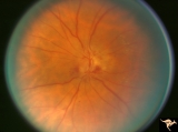
IA09a Evolution of Optociliary Veins with Perioptic Nerve Sheath Meningioma | April 1975, Normal eye, macular degeneration. Anatomy: Optic disc. Pathology: Optociliary vein. Disease/ Diagnosis: Perioptic nerve sheath meningioma evolution. Clinical: Visual loss. | Image |
| 435 |
 |
IA09b Evolution of Optociliary Veins with Perioptic Nerve Sheath Meningioma | January 1977, macular degeneration, disc swelling begins. Anatomy: Optic disc. Pathology: Optociliary vein. Disease/ Diagnosis: Perioptic nerve sheath meningioma evolution. Clinical: Visual loss. | Image |
| 436 |
 |
IA09c Evolution of Optociliary Veins with Perioptic Nerve Sheath Meningioma | June 1977, continued disc swelling. Anatomy: Optic disc. Pathology: Optociliary vein. Disease/ Diagnosis: Perioptic nerve sheath meningioma evolution. Clinical: Visual loss. | Image |
| 437 |
 |
IA09d Evolution of Optociliary Veins with Perioptic Nerve Sheath Meningioma | October 1977, continued disc swelling. Anatomy: Optic disc. Pathology: Optociliary vein. Disease/ Diagnosis: Perioptic nerve sheath meningioma evolution. Clinical: Visual loss. | Image |
| 438 |
 |
IA09e Evolution of Optociliary Veins with Perioptic Nerve Sheath Meningioma | February 1979, development of optociliary veins at 7:00, 1:00. Anatomy: Optic disc. Pathology: Optociliary vein. Disease/ Diagnosis: Perioptic nerve sheath meningioma evolution. Clinical: Visual loss. | Image |
| 439 |
 |
IA09f Evolution of Optociliary Veins with Perioptic Nerve Sheath Meningioma | August 1979, less disc swelling and development of atrophy with more prominent optociliary veins at 7:00 and 1:00. Anatomy: Optic disc. Pathology: Optociliary vein. Disease/ Diagnosis: Perioptic nerve sheath meningioma evolution. Clinical: Visual loss. | Image |
| 440 |
 |
IA09g Evolution of Optociliary Veins with Perioptic Nerve Sheath Meningioma | April 1980, prominent atrophy and increased numbers of optociliary veins. Anatomy: Optic disc. Pathology: Optociliary vein. Disease/ Diagnosis: Perioptic nerve sheath meningioma evolution. Clinical: Visual loss. | Image |
| 441 |
 |
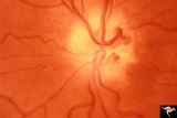
IB101 Post Ischemic (AION) Cupless Atrophy | Left eye, 1993, Male, Post ischemic (AION) cupless atrophy, patient had previous acute AION, Note arteriolar constriction at 7:00 and 11:00, Pairs with IB1_2. Anatomy: Optic disc. Pathology: Post ischemic (AION) cupless atrophy. Disease/ Diagnosis: Post ischemic (AION) cupless atrophy. Clinical: Vis... | Image |
| 442 |
 |
IB102 Post Ischemic (AION) Cupless Atrophy | Right eye, 1993, Male, acute AION. Note the pallid swelling, the small linear hemorrhage at 3:00, and also note that at this stage the retinal arteriole have not become narrowed. Pairs with IB1_1. Anatomy: Optic disc. Pathology: Post ischemic (AION) cupless atrophy. Disease/ Diagnosis: Post ischemic... | Image |
| 443 |
 |
IB103 Post Ischemic (AION) Cupless Atrophy | August 1984, Note severe arteriole narrowing. Anatomy: Optic disc. Pathology: Post ischemic (AION) cupless atrophy. Disease/ Diagnosis: Post ischemic (AION) cupless atrophy. Clinical: Visual loss. | Image |
| 444 |
 |
IB104 Post Ischemic (AION) Cupless Atrophy | January 1969, right eye, striking focal arteriolar narrowing at 4:00 and 10:00. Anatomy: Optic disc. Pathology: Post ischemic (AION) cupless atrophy. Disease/ Diagnosis: Post ischemic (AION) cupless atrophy. Clinical: Visual loss. | Image |
| 445 |
 |
IB105 Post Ischemic (AION) Cupless Atrophy | 1985, right eye, striking focal arteriolar narrowing. Right eye involved two years before the left eye. Woman. Pairs with IB1_6. Anatomy: Optic disc. Pathology: Post ischemic (AION) cupless atrophy. Disease/ Diagnosis: Post ischemic (AION) cupless atrophy. Clinical: Visual loss. | Image |
| 446 |
 |
IB106 Post Ischemic (AION) Cupless Atrophy | 1985, left eye, striking focal arteriolar narrowing. Inferior altitudinal hemianopia. Woman. Pairs with IB1_5. Anatomy: Optic disc. Pathology: Post ischemic (AION) cupless atrophy. Disease/ Diagnosis: Post ischemic (AION) cupless atrophy. Clinical: Visual loss. | Image |
| 447 |
 |
IB107 Post Ischemic (AION) Cupless Atrophy | 1985, right eye, striking focal arteriolar narrowing. Anatomy: Optic disc. Pathology: Post ischemic (AION) cupless atrophy. Disease/ Diagnosis: Post ischemic (AION) cupless atrophy. Clinical: Visual loss. | Image |
| 448 |
 |
IB108 Post Ischemic (AION) Cupless Atrophy | March 1978, right eye, patient had inferior field defect, top half of optic nerve is atrophic, hemiatrophy, blood vessels are focally narrow, bottom half of disc is normal. Anatomy: Optic disc. Pathology: Post ischemic (AION) cupless atrophy. Disease/ Diagnosis: Post ischemic (AION) cupless atrophy.... | Image |
| 449 |
 |
IB109 Post Ischemic (AION) Cupless Atrophy | Right eye, 1983 Top half of disc is pale. Striking focal arteriole narrowing. Anatomy: Optic disc. Pathology: Post ischemic (AION) cupless atrophy. Disease/ Diagnosis: Post ischemic (AION) cupless atrophy. Clinical: Visual loss. | Image |
| 450 |
 |
IB110 Post Ischemic (AION) Cupless Atrophy | Left eye, 1971, upper half of disc is pale with constricted arteriole at 11:00 Anatomy: Optic disc. Pathology: Post ischemic (AION) cupless atrophy. Disease/ Diagnosis: Post ischemic (AION) cupless atrophy. Clinical: Visual loss. | Image |
| 451 |
 |
IB111 Post Ischemic (AION) Cupless Atrophy | Right eye, 1985, patient had inferior altitudinal field defect. Arteriole are narrowing, subtle, but present at 4:00. Anatomy: Optic disc. Pathology: Post ischemic (AION) cupless atrophy. Disease/ Diagnosis: Post ischemic (AION) cupless atrophy. Clinical: Visual loss. | Image |
| 452 |
 |
IB112 Post Ischemic (AION) Cupless Atrophy | Right eye, 1997, pairs with IB1_13, striking arteriole narrowing. Anatomy: Optic disc. Pathology: Post ischemic (AION) cupless atrophy. Disease/ Diagnosis: Post ischemic (AION) cupless atrophy. Clinical: Visual loss. | Image |
| 453 |
 |
IB113 Post Ischemic (AION) Cupless Atrophy | Left eye, 1997, pairs with IB1_12, striking arteriole narrowing. Anatomy: Optic disc. Pathology: Post ischemic (AION) cupless atrophy. Disease/ Diagnosis: Post ischemic (AION) cupless atrophy. Clinical: Visual loss. | Image |
| 454 |
 |
IB114a Post Ischemic (AION) Cupless Atrophy | 1991, acute AION in a disc with a cup, pair with IB1_14b. Anatomy: Optic disc. Pathology: Post ischemic (AION) cupless atrophy. Disease/ Diagnosis: Post ischemic (AION) cupless atrophy. Clinical: Visual loss. | Image |
| 455 |
 |
IB114b Post Ischemic (AION) Atrophy in a Disc with a Cup | 1996, same as IB1_14a five years later reveals pallor, arteriole narrowing and optic cup. Anatomy: Optic disc. Pathology: Post ischemic (AION) cupless atrophy. Disease/ Diagnosis: Post ischemic (AION) cupless atrophy. Clinical: Visual loss. | Image |
| 456 |
 |
IB201 Post Radiation Papillopathy | Right eye, 1982, Bizarre drusen like bodies on the pale atrophic disc. Note arteriolar narrowing. Note peripapillary circumferential retinal exudate. Anatomy: Optic disc. Pathology: Post radiation papillopathy. Disease/ Diagnosis: Post radiation papillopathy. Clinical: Blindness following radiation ... | Image |
| 457 |
 |
IB202 Post Radiation Papillopathy | Left eye, 1987, Marked arteriolar narrowing. Occluded arteriole at 7:00 Diffuse hemorrhage. Neovascularization on the disc. Anatomy: Optic disc. Pathology: Post radiation papillopathy. Disease/ Diagnosis: Post radiation papillopathy. Clinical: Blindness following radiation therapy for tumors | Image |
| 458 |
 |
IB203 Post Radiation Papillopathy | 1975, left eye. Bizarre arteriolar narrowings and neovascularization. Anatomy: Optic disc. Pathology: Post radiation papillopathy. Disease/ Diagnosis: Post radiation papillopathy. Clinical: Blindness following radiation therapy for tumors. | Image |
| 459 |
 |
IB204 Post Radiation Papillopathy | 1973. Although patient lived, she was blinded by her radiation treatment for glioma. Note retinal arterioles are so small they are barely visible. Anatomy: Optic disc. Pathology: Post radiation papillopathy. Disease/ Diagnosis: Post radiation papillopathy. Clinical: Blindness following radiation the... | Image |
| 460 |
 |
IC101 Old Central Retinal Artery Occlusion | Old central retinal artery occlusion without additional retinovascular signs, 1969. Anatomy: Optic disc. Pathology: Central retinal artery occlusion. Disease/ Diagnosis: Central retinal artery occlusion. Clinical: Sudden blindness. | Image |
| 461 |
 |
IC102a Central Retinal Artery Occlusion with Cilioretinal Collaterals | Left eye, 1988, Central retinal artery with cilioretinal collaterals due to calcific embolic behind the lamina cribrosa due to calcific valvular heart disease. Collaterals have been called "Nettleship Collaterals", recognizing the British physician who first described them in 1892. Anatomy: Optic di... | Image |
| 462 |
 |
IC102b Central Retinal Artery Occlusion with Cilioretinal Collaterals | Right eye, 1991, Central retinal artery occlusion with cilioretinal collateral occlusions due to calcific embolic occlusion behind the lamina cribrosa due to calcific valvular heart disease. Collaterals have been called "Nettleship Collaterals", recognizing the British physician who first described ... | Image |
| 463 |
 |
IC102c Central Retinal Artery Occlusion with Cilioretinal Collaterals | Right eye, 1982, Central retinal artery occlusion with cilioretinal collateral occlusions due to calcific embolic occlusion behind the lamina cribrosa due to calcific valvular heart disease. Collaterals have been called "Nettleship Collaterals", recognizing the British physician who first described ... | Image |
| 464 |
 |
IC103a Central Retinal Artery Occlusion with Choroidal Arteriolar Occlusion | Central retinal artery occlusion and choroidal vascular occlusion due to pressure on the eyeball during craniotomy. Note total loss of vascularity of the optic disc and surrounding choroid. Anatomy: Optic disc. Pathology: Combined central retinal and choroidal arteriolar occlusion. Disease/ Diagnos... | Image |
| 465 |
 |
IC103b Central Retinal Artery Occlusion with Choroidal Arteriole Occlusion | 1980, Evidence of choroidal vascular ischemia. Central retinal artery occlusion and choroidal vascular occlusion from amputation of the optic nerve for meningioma. Anatomy: Optic disc. Pathology: Optic glioma and optic nerve was amputated during excision of the tumor. Disease/ Diagnosis: Optic nerve... | Image |
| 466 |
 |
IC103c Central Retinal Artery Occlusion with Choroidal Arteriole Occlusion | 1988, Central retinal artery occlusion and choroidal vascular occlusion, 70 year old woman with history of central retinal artery occlusion 30 years prior. Anatomy: Optic disc. Pathology: Combined central retinal and choroidal arteriolar occlusion. Disease/ Diagnosis: Combined central retinal and ch... | Image |
| 467 |
 |
IC103d Central Retinal Artery Occlusion with Choroidal Arteriole Occlusion | 1969, Complete loss of blood supply to retina and choroid. Cause unknown. Boy. Anatomy: Optic disc. Pathology: Toxic ischemic retinal damage 22a. Clinical: Blindness. | Image |
| 468 |
 |
IC104a Retinal Pigmentary Degeneration (Sine Pigmentosa) | Retinal pigmentary degeneration (sine pigmentosa), with extreme arteriolar narrowing. 1967. Anatomy: Optic disc. Pathology: Retinal pigmentary degeneration. Disease/ Diagnosis: Retinal pigmentary degeneration. Clinical: Night blindness. | Image |
| 469 |
 |
IC105 Quinine Toxicity (Amblyopia) | Quinine toxicity (amblyopia), 1971, blind eye, note diffuse arteriole narrowing and optic nerve pallor. Anatomy: Optic disc. Pathology: Toxic ischemic retinal damage. Disease/ Diagnosis: Quinine toxicity. Clinical: Blindness. | Image |
| 470 |
 |
ID01 Post Papilledema Gliosis | Post papilledema milky gliosis with arteriolar constriction, 1982, right eye, pair with ID_2. Anatomy: Optic disc. Pathology: Post papilledema atrophy and gliosis due to huge anterior communicating artery aneurysm. Disease/ Diagnosis: Elevated intracranial pressure from aneurysm. Clinical: Diminishe... | Image |
| 471 |
 |
ID02 Post Papilledema Gliosis | Post papilledema milky gliosis with arteriolar constriction and atrophy, 1982, left eye, pair with ID_1. Anatomy: Optic disc. Pathology: Post papilledema atrophy and gliosis due to huge anterior communicating artery aneurysm. Disease/ Diagnosis: Elevated intracranial pressure from aneurysm. Clinical... | Image |
| 472 |
 |
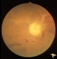
ID03a Post Papilledema Atrophy with Marked Gliosis | Post papilledema atrophy with marked gliosis in a patient with pseudotumor cerebri. Patient weighed over 300 pounds. Right eye blind. 1981. Right eye. Pair with ID_3b. Anatomy: Optic disc. Pathology: Post papilledema atrophy and gliosis from long standing elevated intracranial pressure. Disease/ Dia... | Image |
| 473 |
 |
ID03b Post Papilledema Atrophy with Marked Gliosis | Post papilledema atrophy with marked gliosis in a patient with pseudotumor cerebri. Patient weighed over 300 pounds. Left eye has visual field defects. 1981, right eye, pair with ID_3a. Anatomy: Optic disc. Pathology: Post papilledema atrophy and gliosis from long standing elevated intracranial pres... | Image |
| 474 |
 |
ID04a Post Papilledema Atrophy with Marked Gliosis | Post papilledema atrophy with marked gliosis in a patient with pseudotumor cerebri, 1985, right eye, pair with ID_4b, Note "high water" marks in peripapillary pigment epithelial layer. Anatomy: Optic disc. Pathology: Post papilledema atrophy and gliosis from long standing elevated intracranial press... | Image |
| 475 |
 |
ID04b Post Papilledema Atrophy with Marked Gliosis | Post papilledema atrophy with marked gliosis in a patient with pseudotumor. Nasal ovoid absence of the retinal pigment epithelium. Presumably a defect from the long standing papilledema. 1985,. Right eye, pair with ID_4a. Anatomy: Optic disc. Pathology: Post papilledema atrophy and gliosis from long... | Image |
| 476 |
 |
ID05a Post Papilledema Optic Atrophy from Pseudotumor Cerebri | Left eye, October 1999, Post papilledema optic atrophy from pseudotumor cerebri. Note optociliary veins in both discs. Gliosis and partial pallor following long standing papilledema and intracranial pressure. Anatomy: Optic disc. Pathology: Post papilledema atrophy and gliosis from long standing el... | Image |
| 477 |
 |
ID05b Post Papilledema Optic Atrophy from Pseudotumor Cerebri | Right eye, October 1999, Post papilledema optic atrophy from pseudotumor cerebri. Note optociliary veins in both discs. Gliosis and partial pallor following long standing papilledema and intracranial pressure. Anatomy: Optic disc. Pathology: Post papilledema atrophy and gliosis from long standing el... | Image |
| 478 |
 |
ID06 Post Papilledema Optic Atrophy with Arteriolar Sheathing and Optociliary Veins | 1989. Post papilledema optic atrophy with arteriolar sheathing and optociliary veins. Anatomy: Optic disc. Pathology: Long standing effects of intracranial pressure. Clinical: Blindness. | Image |
| 479 |
 |
ID07 Post Papilledema Optic Atrophy | Post papilledema optic atrophy with gliosis and arteriolar narrowing. 1994. Anatomy: Optic disc. Pathology: Residue of long standing papilledema. Clinical: Visual loss. | Image |
| 480 |
 |
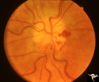
IE01 Acute Leber Optic Neuropathy | Pseudo edema with peripapillary microangiopathy in a brother of boy with Leber Optic Neuropathy. Not pale nerve yet. Right eye. Same patient as IE_02a and IE_02b. August 7, 1979. Anatomy: Optic disc. Pathology: Optic neuropathy. Disease/ Diagnosis: Optic neuropathy. Clinical: Asymptomatic | Image |
| 481 |
 |
IE02a Acute Leber Optic Neuropathy | Acute Leber Optic Neuropathy, Left eye. Same patient as IE_01 and IE_02b. August 7, 1979. Anatomy: Optic disc. Pathology: Optic neuropathy. Disease/ Diagnosis: Leber's optic neuropathy. Clinical: Asymptomatic. | Image |
| 482 |
 |
IE02b Acute Leber Optic Neuropathy | Acute Leber Optic Neuropathy. Formation of hemorrhage one year after IE_02a. At this time, he was beginning to lose central vision. .Note thinning of the nerve fiber layer temporally, 3:00 - 4:00 Pair with IE_02a. March 26, 1980. Anatomy: Optic disc. Pathology: Optic neuropathy. Disease/ Diagnosis: ... | Image |
| 483 |
 |
IE03 Acute Leber Optic Neuropathy | Acute stage of Leber optic neuropathy with microangiopathy and peripapillary nerve fiber layer thickening. The temporal nerve fiber layer is already showing atrophy. Central vision is grossly reduced. 1971. Anatomy: Optic disc. Pathology: Optic neuropathy. Disease/ Diagnosis: Leber's optic neuropath... | Image |
| 484 |
 |
IE04 Acute Leber Optic Neuropathy | Microangiopathy without visual loss in a patient with acute Leber's optic neuropathy in the left eye. Pair with IE_05. Anatomy: Optic disc. Pathology: Optic neuropathy. Disease/ Diagnosis: Leber's optic neuropathy. Clinical: Central vision loss. | Image |
| 485 |
 |
IE05 Acute Leber Optic Neuropathy | Patient has just begun to lose vision in his left eye due to Leber's optic neuropathy. Pair with IE_04. Anatomy: Optic disc. Pathology: Optic neuropathy. Disease/ Diagnosis: Leber's optic neuropathy. Clinical: Central vision loss. | Image |
| 486 |
 |
IE06 Subacute Leber Optic Neuropathy | Subacute Leber's optic neuropathy with microangiopathy with distinct temporal disc pallor. 1971. Anatomy: Optic disc. Pathology: Optic neuropathy. Disease/ Diagnosis: Leber's optic neuropathy. Clinical: Large central vision loss. | Image |
| 487 |
 |
IE07 Subacute Leber Optic Neuropathy | Subacute Leber's optic neuropathy with microangiopathy. 1973. Anatomy: Optic disc. Pathology: Optic neuropathy. Disease/ Diagnosis: Leber's optic neuropathy. Clinical: Early central vision loss. | Image |
| 488 |
 |
IE08a Subacute Leber Optic Neuropathy | Subacute Leber Optic Neuropathy with temporal atrophy. August 5, 1980. Pair with IE_1, 2a&b, IE_8b, IE_9a&b. Anatomy: Optic disc. Pathology: Optic neuropathy. Disease/ Diagnosis: Leber's optic neuropathy. Clinical: Visual loss. | Image |
| 489 |
 |
IE08b Subacute Leber Optic Neuropathy | Subacute Leber Optic Neuropathy with temporal atrophy. August,1980. Pair with IE_1, 2a&b, IE_8a, IE_9a&b. Anatomy: Optic disc. Pathology: Optic neuropathy. Disease/ Diagnosis: Leber's optic neuropathy. Clinical: Visual loss. | Image |
| 490 |
 |
IE09a Chronic Leber Optic Neuropathy | Chronic Leber Optic Neuropathy with advancing temporal pallor. Notice the nerve fiber layer thickening has diminished. November 13, 1980. Pair with IE_1, 2a&b, IE_9b, IE_8a&b. Anatomy: Optic disc. Pathology: Optic neuropathy. Disease/ Diagnosis: Leber's optic neuropathy. Clinical: Blindness. | Image |
| 491 |
 |
IE09b Chronic Leber Optic Neuropathy | Chronic Leber Optic Neuropathy with advancing temporal pallor. Notice the nerve fiber layer thickening has diminished. November 13, 1980. Pair with IE_1, 2a&b, IE_9a, IE_8a&b. Anatomy: Optic disc. Pathology: Optic neuropathy. Disease/ Diagnosis: Leber's optic neuropathy. Clinical: Blindness. | Image |
| 492 |
 |
IE10a Chronic Leber Optic Neuropathy | Chronic Leber's Optic Neuropathy, August 8, 1969. Anatomy: Optic disc. Pathology: Optic neuropathy. Disease/ Diagnosis: Leber's optic neuropathy. Clinical: Blindness. | Image |
| 493 |
 |
IE10b Subacute Leber Optic Neuropathy | Subacute Leber's Optic Neuropathy, August 8, 1969, Left eye, pair with IE_10a, c. Anatomy: Optic disc. Pathology: Optic neuropathy. Disease/ Diagnosis: Leber's optic neuropathy. Clinical: Blindness. | Image |
| 494 |
 |
IE10c Chronic Leber Optic Neuropathy | February 12, 1970, Chronic Leber's Optic Neuropathy, 6 month follow up from 10b. Thickening of the nerve fiber layer is gone. Left eye, pair with IE_10a, c. Anatomy: Optic disc. Pathology: Optic neuropathy. Disease/ Diagnosis: Leber's optic neuropathy. Clinical: Blindness. | Image |
| 495 |
 |
IE11 Subacute Leber Optic Neuropathy | Subacute Leber's Optic Neuropathy with distinct temporal wedge pallor and adjacent microangiopathy. 1973. Anatomy: Optic disc. Pathology: Optic neuropathy. Disease/ Diagnosis: Leber's optic neuropathy. Clinical: Blindness. | Image |
| 496 |
 |
IE12a Acute Leber Optic Neuropathy | Subacute stage of Leber's Optic Neuropathy showing microangiopathy showing temporal pallor. Atrophy more advanced in left eye (12b) 1971, right eye. Anatomy: Optic disc. Pathology: Optic neuropathy. Disease/ Diagnosis: Leber's optic neuropathy. Clinical: Blindness. | Image |
| 497 |
 |
IE12b Subacute Leber Optic Neuropathy | Subacute stage of Leber's Optic Neuropathy showing microangiopathy showing temporal pallor. Atrophy more advanced in left eye. 1971, left eye. Anatomy: Optic disc. Pathology: Optic neuropathy. Disease/ Diagnosis: Leber's optic neuropathy. Clinical: Blindness. | Image |
| 498 |
 |
IE13a End Stage Leber Optic Neuropathy | End stage Leber's Optic Neuropathy. Note modest arteriolar narrowing. Note also the generalized pallor of the disc. Microangiopathy is no longer visible. Right eye. Pair with 13b. Anatomy: Optic disc. Pathology: Optic neuropathy. Disease/ Diagnosis: Leber's optic neuropathy. Clinical: Blindness. | Image |
| 499 |
 |
IE13b End Stage Leber Optic Neuropathy | End stage Leber's Optic Neuropathy. Note modest arteriolar narrowing. Note also the generalized pallor of the disc. Microangiopathy is no longer visible. Left eye. Pair with 13a. Anatomy: Optic disc. Pathology: Optic neuropathy. Disease/ Diagnosis: Leber's optic neuropathy. Clinical: Blindness. | Image |
| 500 |
 |
IE14a End Stage Leber Optic Neuropathy | End stage Leber's Optic Neuropathy. Dense temporal pallor. Microangiopathy is absent. Right eye. Pair with 14b. Anatomy: Optic disc. Pathology: Optic neuropathy. Disease/ Diagnosis: Leber's optic neuropathy. Clinical: Blindness. | Image |