Best known for his world-renowned neuro-ophthalmology unit based at the University of California, San Francisco, William Hoyt, MD collected here more than 850 of his best images covering a wide range of disorders.
William F. Hoyt, MD, Professor Emeritus of Ophthalmology, Neurology and Neurosurgery, Department of Ophthalmology, University of California, San Francisco.
NOVEL: https://novel.utah.edu/
TO
| Title | Description | Type | ||
|---|---|---|---|---|
| 76 |
 |
C308 Nodular Papillopathies (Sarcoid) | Nodular infiltrative papillopathy in a patient with sarcoid. Woman. Anatomy: Optic disc. Pathology: Axoplasmic stasis due to sarcoid infiltration. Disease/ Diagnosis: Sarcoid papillopahty. Clinical: Unknown? | Image |
| 77 |
 |
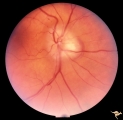
C37 Anomalous Pale Disc | "Watermelon" disc. Woman. Normal function. Left eye. Anatomy: Optic disc. | Image |
| 78 |
 |
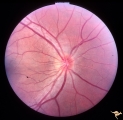
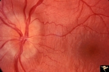
C401 Luetic Papillopathy (Gumma of the Optic Disc) | Diffuse optic disc swelling with tortuous capillary dilations indicating inflammatory cellular infiltration. October 2001. Same eye as C4_02. Anatomy: Optic disc. Pathology: Axoplasmic stasis due to syphillitic infection. Luetic papillopathy (Syphyllis). Clinical: Visual loss. | Image |
| 79 |
 |
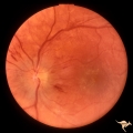
C402 Luetic Papillopathy (Gumma of the Optic Disc) | November 2001. Same eye as C4_01 after treatment with penicillin. Disc swelling went away and good visual function returned. Anatomy: Optic disc. Pathology: Axoplasmic stasis due to syphillitic infection. Disease/ Diagnosis: Luetic papillopathy (Syphillis). Clinical: Improving visual loss. | Image |
| 80 |
 |
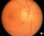
Cerebellar Macular Degenerative Disease | Ocular fundus shows prominent retinal degeneration in the region of the maculae, bilateral optic disc pallor with narrowed retinal arterioles. Interesting peripapillary halo of retinal pigment degeneration. Most consistent with Spinal Cerebellar Degeneration Type 7 (SCA-7). Anatomy: Retina. Patholog... | Image |
| 81 |
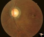 |
Cerebellar Macular Degenerative Disease | Ocular fundus shows prominent retinal degeneration in the region of the maculae, bilateral optic disc pallor with narrowed retinal arterioles. Interesting peripapillary halo of retinal pigment degeneration. Most consistent with Spinal Cerebellar Degeneration Type 7 (SCA-7). Anatomy: Retina. Patholog... | Image |
| 82 |
 |
Cerebroretinal Microangiopathy (Susac Syndrome) | Two plaques which have been called Psuedo-emboli. This plaque is not the result of embolism, but is the result of the microangioplastic process underlying the syndrome. (NANOS 2001 by Egan, RA). Anatomy: Retina. Pathology: Microangiopathy involving brain, auditory nerve and retina. Disease/Diagnosi... | Image |
| 83 |
 |
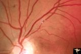
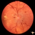
Cerebroretinal Microangiopathy (Susac Syndrome) | Retinal signs of Susac's Syndrome in acute phase consist of areas of retinal artery infarction from branch retinal artery occlusions. This fundus shows two area of retinal infarction from occlusion of both superior and inferior branch retinal arterioles. Anatomy: Retina. Pathology: Microangiopathy... | Image |
| 84 |
 |
Cerebroretinal Microangiopathy (Susac Syndrome) | Retinal signs of Susac's Syndrome in acute phase consist of areas of retinal artery infarction from branch retinal artery occlusions. These patients are usually women, many of whom are demented and have hearing loss. Refs: 1) Susac, Hardiman, Sellhorst. Neurology. 1979. 29:313-316 2) Susac ""Susa... | Image |
| 85 |
 |
Cerebroretinal Microangiopathy (Susac Syndrome) | This fundus picture from a patient with Susac Syndrome shows a focal shiny plaque in the inferior retinal arteriole. This plaque is not the result of embolism, but is the result of the microangioplastic process underlying the syndrome. (NANOS 2001 by Egan, RA). Anatomy: Retina. Pathology: Microangi... | Image |
| 86 |
 |
Cerebroretinal Microangiopathy (Susac Syndrome) | Retinal signs of Susac's Syndrome in acute phase consist of areas of retinal artery infarction from branch retinal artery occlusions. Shows clearing retinal branch artery occlusion. Pathology: Retina. Pathology: Microangiopathy involving brain, auditory nerve and retina. Disease/Diagnosis: Cerebro... | Image |
| 87 |
 |
Cerebroretinal Microangiopathy (Susac Syndrome) | Retinal signs of Susac's Syndrome in acute phase consist of areas of retinal artery infarction from branch retinal artery occlusions. Branch artery occlusion beginning to clear. Note the occluded arteriole lying on top of the infarcted zone. Anatomy: Retina. Pathology: Microangiopathy involving br... | Image |
| 88 |
 |
Cerebroretinal Microangiopathy (Susac Syndrome) | There is a plaque superior to the disc at 12:00. This plaque is not the result of embolism, but is the result of the microangioplastic process underlying the syndrome. There is a ghost vessel inferiorly at 5:00 off the disc. (NANOS 2001 by Egan, RA). Anatomy: Retina. Pathology: Microangiopathy invo... | Image |
| 89 |
 |
Cerebroretinal Microangiopathy (Susac Syndrome) | There is an occlusion of the superior nasal retinal arteriole visible as a white ghost vessel at 11:00. Note: Collateral filling of the distal branches in two places. (NANOS 2001 by Egan, RA). Anatomy: Retina. Pathology: Microangiopathy involving brain, auditory nerve and retina. Disease/Diagnosis:... | Image |
| 90 |
 |
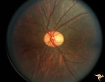
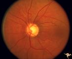
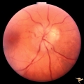
Congenitally Crowded Disc - Little Red Disc | Right eye: "little red disc". Congenitally blurred disc. 26 year old man. Anatomy: Optic disc Pathology: Normal variation of the optic disc Disease/Diagnosis: Normal variation of the optic disc. Congenital blurred disc. Little red disc. | Image |
| 91 |
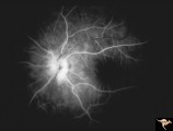 |
D106 Disc Edema with Systemic Lupus | Flourescein angiogram shows evidence of vascular papillopathy. (Lupus) Same patient as D1_05 an D1_07. Anatomy: Optic disc. Pathology: Axoplasmic stasis due to vasculitis (Lupus). Disease/ Diagnosis: Lupus papillopathy. | Image |
| 92 |
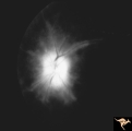 |
D107 Disc Edema with Systemic Lupus | Late stage Flourescein angiogram showing flourescein leakage on the disc and around the neighboring vessels. Note this amount of edema could not be appreciated in the colored fundus image D1_05. Same patient as D1_06 an D1_05. Anatomy: Optic disc. Pathology: Axoplasmic stasis due to vasculitis (Lupu... | Image |
| 93 |
 |
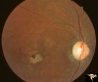
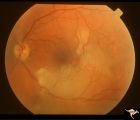
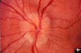
D201 Disc Edema with Systemic Hypertension | Left eye. Note generalized arterial narrowing. Low grade disc edema and multiple splinter hemorrhages. The patient had severe hypertension from kidney failure. Additional yellow intraretinal exudate at the macula. 20 year old male patient. Right eye. Pair with D2_02. Anatomy: Optic disc; Retina; Ret... | Image |
| 94 |
 |
D202 Disc Edema with Systemic Hypertension | Right eye. Note generalized arteriole narrowing. Low grade disc edema and multiple splinter hemorrhages. The patient had severe hypertension from kidney failure. Additional yellow intraretinal exudate at the macula. 20 year old male patient. Pair with D2_01. Anatomy: Optic disc; Retina; Retinal art... | Image |
| 95 |
 |
Drusen Plus Papilledema | PP37a: right swollen disc on top of drusen with narrowing of the arterioles; PP37b: left visible drusen and papilledema with sub-retinal hemorrhage temporally. Patient had frontal glioblastoma. Anatomy: Optic disc. Pathology: Drusen of the optic disc. Disease/Diagnosis: Drusen of the optic disc. Cli... | Image |
| 96 |
 |
Drusen Plus Papilledema | PP37a: right swollen disc on top of drusen with narrowing of the arterioles;PP37 b: left visible drusen and papilledema with sub-retinal hemorrhage temporally. Patient had frontal glioblastoma. Anatomy: Optic disc. Pathology: Drusen of the optic disc. Disease/Diagnosis: Drusen of the optic disc. Cli... | Image |
| 97 |
 |
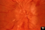
Drusen with Horizontal Retinal Folds | PP35a: Right eye. Buried drusen. PP35b: Left eye. Buried drusen with retinal folds. 21 year old woman. Anatomy: Optic disc. Pathology: Drusen of the optic disc. Disease/Diagnosis: Drusen of the optic disc. | Image |
| 98 |
 |
Drusen with Horizontal Retinal Folds | PP35: Right eye. Buried drusen. PP35b: Left eye. Buried drusen with retinal folds. 21 year old woman. Anatomy: Optic disc. Pathology: Drusen of the optic disc. Disease/Diagnosis: Drusen of the optic disc. | Image |
| 99 |
 |
Drusen with Vertical Retinal Folds | PP36a & b: Both left eye: Buried drusen. Note vertical retinal folds. Anatomy: Optic disc. Pathology: Drusen of the optic disc. Disease/Diagnosis: Drusen of the optic disc. | Image |
| 100 |
 |
Drusen with Vertical Retinal Folds | PP36a & b:Both left eye: Buried drusen. Note vertical retinal folds. Anatomy: Optic disc. Pathology: Drusen of the optic disc. Disease/Diagnosis: Drusen of the optic disc. | Image |