Best known for his world-renowned neuro-ophthalmology unit based at the University of California, San Francisco, William Hoyt, MD collected here more than 850 of his best images covering a wide range of disorders.
William F. Hoyt, MD, Professor Emeritus of Ophthalmology, Neurology and Neurosurgery, Department of Ophthalmology, University of California, San Francisco.
NOVEL: https://novel.utah.edu/
TO
Filters: Collection: "ehsl_novel_wfh"
| Title | Description | Type | ||
|---|---|---|---|---|
| 76 |
 |
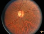
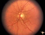
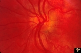
H61 Superior Segmental Optic Hypoplasia (SSOH) Topless Disc Syndrome | Left eye. SSOH. Same patient as H_62 and H_63. Anatomy: Optic disc. Pathology: Superior segmental optic hypoplasia (SSOH). Disease/ Diagnosis: Congenital anomaly. | Image |
| 77 |
 |
H63 Superior Segmental Optic Hypoplasia (SSOH) Topless Disc Syndrome | Visual fields. Right eye has mild depression. Same patient as H_61 an H_62. Anatomy: Optic disc. Pathology: Superior segmental optic hypoplasia (SSOH). Disease/ Diagnosis: Congenital anomaly. | Image |
| 78 |
 |
H64 Superior Segmental Optic Hypoplasia (SSOH) Topless Disc Syndrome | Bilateral SSOH. Same patient as H_65 and H_66. Anatomy: Optic disc. Pathology: Superior segmental optic hypoplasia (SSOH). Disease/ Diagnosis: Congenital anomaly. | Image |
| 79 |
 |
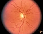
H65 Superior Segmental Optic Hypoplasia (SSOH) Topless Disc Syndrome | Bilateral SSOH. Same patient as H_64 and H_66. Anatomy: Optic disc. Pathology: Superior segmental optic hypoplasia (SSOH). Disease/ Diagnosis: Congenital anomaly. | Image |
| 80 |
 |
H67 Superior Segmental Optic Hypoplasia (SSOH) Topless Disc Syndrome | Bilateral SSOH. Right eye. Same patient as H_68 and H_69. Anatomy: Optic disc. Pathology: Superior segmental optic hypoplasia (SSOH). Disease/ Diagnosis: Congenital anomaly. | Image |
| 81 |
 |
H70 Superior Segmental Optic Hypoplasia (SSOH) Topless Disc Syndrome | Bilateral SSOH. Right eye. Same patient as H_71 and H_72. Anatomy: Optic disc. Pathology: Superior segmental optic hypoplasia (SSOH). Disease/ Diagnosis: Congenital anomaly. | Image |
| 82 |
 |
H71 Superior Segmental Optic Hypoplasia (SSOH) Topless Disc Syndrome | Bilateral SSOH. Left eye. Same patient as H_70 and H_72. Anatomy: Optic disc. Pathology: Superior segmental optic hypoplasia (SSOH). Disease/ Diagnosis: Congenital anomaly. | Image |
| 83 |
 |
H73 Superior Segmental Optic Hypoplasia (SSOH) Topless Disc Syndrome | Bilateral SSOH. Left eye. Same patient as H_73. Anatomy: Optic disc. Pathology: Superior segmental optic hypoplasia (SSOH). Disease/ Diagnosis: Congenital anomaly. | Image |
| 84 |
 |
H74 Superior Segmental Optic Hypoplasia (SSOH) Topless Disc Syndrome | Bilateral SSOH. Right eye. Same patient as H_74. Anatomy: Optic disc. Pathology: Superior segmental optic hypoplasia (SSOH). Disease/ Diagnosis: Congenital anomaly. | Image |
| 85 |
 |
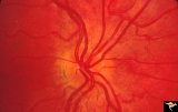
H75 Superior Segmental Optic Hypoplasia (SSOH) Topless Disc Syndrome | SSOH with hypoplasia of whole disc. Right eye. Anatomy: Optic disc. Pathology: Superior segmental optic hypoplasia (SSOH). Disease/ Diagnosis: Congenital anomaly. | Image |
| 86 |
 |
H76 Superior Segmental Optic Hypoplasia (SSOH) Topless Disc Syndrome | SSOH. Right eye. Anatomy: Optic disc. Pathology: Superior segmental optic hypoplasia (SSOH). Disease/ Diagnosis: Congenital anomaly. | Image |
| 87 |
 |
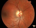
H95 Occipital Hemianoptic Hypoplasia | Right eye with temporal field defect shows trans-synaptic band atrophy. Absence of nasal nerve fibers. Same patient as H_93, H_94, H_96, H_97. Anatomy: Optic disc. Pathology: Occipital hemianoptic hypoplasia. Disease/ Diagnosis: Congenital defect of the occipital lobe. Imaging: MRI scan - See slide ... | Image |
| 88 |
 |
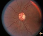
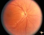
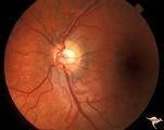
H58 Superior Segmental Optic Hypoplasia (SSOH) Topless Disc Syndrome | Right eye. Same patient as H_59 and H_60. Original case identifying SSOH, or "Topless disc syndrome" from Zurich. Anatomy: Optic disc. Pathology: Superior segmental optic hypoplasia (SSOH). Disease/ Diagnosis: Congenital anomaly. | Image |
| 89 |
 |
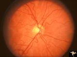
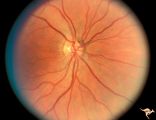
H59 Superior Segmental Optic Hypoplasia (SSOH) Topless Disc Syndrome | Left eye. Same patient as H_58 and H_60. Original case identifying SSOH, or "Topless disc syndrome" from Zurich. Anatomy: Optic disc. Pathology: Superior segmental optic hypoplasia (SSOH). Disease/ Diagnosis: Congenital anomaly. | Image |
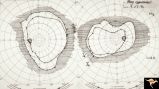
| 90 |
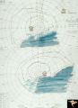 |
H60 Superior Segmental Optic Hypoplasia (SSOH) Topless Disc Syndrome | Visual fields. Bilateral inferior altitudinal field defects. Same patient as H_59 and H_58. Original case identifying SSOH, or "Topless disc syndrome" from Zurich. Anatomy: Optic disc. Pathology: Superior segmental optic hypoplasia (SSOH). Disease/ Diagnosis: Congenital anomaly. | Image |
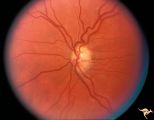
| 91 |
 |
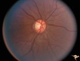
H62 Superior Segmental Optic Hypoplasia (SSOH) Topless Disc Syndrome | Right eye. Disc looks almost normal but superior nerve fiber layer is thinned and represents a mild form of SSOH. Same patient as H_61 and H_63. Anatomy: Optic disc. Pathology: Superior segmental optic hypoplasia (SSOH). Disease/ Diagnosis: Congenital anomaly. | Image |
| 92 |
 |
H68 Superior Segmental Optic Hypoplasia (SSOH) Topless Disc Syndrome | Bilateral SSOH. Left eye. Entire nerve looks hypoplastic. Same patient as H_67 and H_69. Anatomy: Optic disc. Pathology: Superior segmental optic hypoplasia (SSOH). Disease/ Diagnosis: Congenital anomaly. | Image |
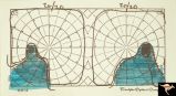
| 93 |
 |
H69 Superior Segmental Optic Hypoplasia (SSOH) Topless Disc Syndrome | Visual field. Entire visual field is constricted. Patient's mother Type 1 diabetic. Same patient as H_68 and H_69. Anatomy: Optic disc. Pathology: Superior segmental optic hypoplasia (SSOH). Disease/ Diagnosis: Congenital anomaly. | Image |
| 94 |
 |
H72 Superior Segmental Optic Hypoplasia (SSOH) Topless Disc Syndrome | Visual field. Entire visual field is constricted. Patient's mother Type 1 diabetic. Same patient as H_70 and H_71. Anatomy: Optic disc. Pathology: Superior segmental optic hypoplasia (SSOH). Disease/ Diagnosis: Congenital anomaly. | Image |
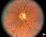
| 95 |
 |
H52 Superior Segmental Optic Hypoplasia (SSOH) Topless Disc Syndrome | High exit point of central retinal vessels. Superior choroidal crescent. Complete loss of nerve fiber layer entering disc from above. Inferior altitudinal field defect. Type 1 diabetic mother. Anatomy: Optic disc. Pathology: Superior segmental optic hypoplasia (SSOH). Disease/ Diagnosis: Congenital ... | Image |
| 96 |
 |
Bilateral Crowded Discs | Left eye. Bilateral crowded discs with congenital blurring. Blurred disc margins are not from edema. Note optic cup is absent. Pair with right eye in PP_1a, and brother in PP_2. Mother has drusen of the optic disc in PP_11aa & b. Sister has drusen in PP_11c. Anatomy: Optic disc. Pathology: Normal va... | Image |
| 97 |
 |
Bilateral Crowded Discs (Family) | Right eye. Bilateral crowded discs with congenital blurring. Blurred disc margins are not from edema. Note optic cup is absent. Pair with left eye in PP_1b, and brother in PP_2. Mother has drusen of the optic disc in PP_11a & b. Sister has drusen in PP_11c. Anatomy: Optic disc. Pathology: Normal var... | Image |
| 98 |
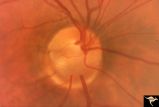 |
C01 Pits of the Optic Disc | Right eye. Very large inferior temporal optic pit. Congenital. Woman. Anatomy: Optic disc. | Image |
| 99 |
 |
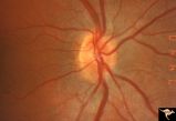
C36 Anomalous Pale Disc | Multiple cilioretinal arteries. Pale appearance. Normal optic nerve function. Good example of "empty disc". Pair with C_33. Anatomy: Optic disc. | Image |
| 100 |
 |
H04 Panhypoplasia | Right eye. Normal eye. Girl. Same patient as H_3. Anatomy: Optic disc. Pathology: Hypoplasia of the optic nerve. Disease/ Diagnosis: Hypoplasia. | Image |