Best known for his world-renowned neuro-ophthalmology unit based at the University of California, San Francisco, William Hoyt, MD collected here more than 850 of his best images covering a wide range of disorders.
William F. Hoyt, MD, Professor Emeritus of Ophthalmology, Neurology and Neurosurgery, Department of Ophthalmology, University of California, San Francisco.
NOVEL: https://novel.utah.edu/
TO
Filters: Collection: "ehsl_novel_wfh"
| Title | Description | Type | ||
|---|---|---|---|---|
| 51 |
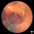 |
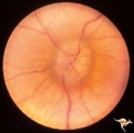
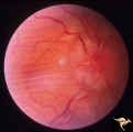
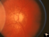
Chronic Papilledema with Hemorrhagic and Exudative Complications | Left eye at presesntation. Chronic papilledema with hemorrhagic and exudative complications due to Pseudotumor cerebri. Anatomy: Optic disc Pathology: Papilledema Disease/Diagnosis: Chronic papilledema with hemorrhagic and exudative complications. | Image |
| 52 |
 |
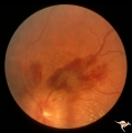
Chronic Papilledema with Hemorrhagic and Exudative Complications | Left eye one month after presentation. View below of resolving subretinal hemorrhage. Chronic papilledema with hemorrhagic and exudative complications due to Pseudotumor cerebri. | Image |
| 53 |
 |
Chronic Papilledema with Hemorrhagic and Exudative Complications | Left eye one month after presentation. View above of resolving preretinal hemorrhage. Chronic papilledema with hemorrhagic and exudative complications due to Pseudotumor cerebri. | Image |
| 54 |
 |
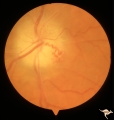
Chronic Papilledema with Pseudo Drusen | Left eye. Chronic papilledema with pseudo drusen due to cerebral pontine angle tumor. Anatomy: Optic disc. Pathology: Papilledema Disease/Diagnosis: Chronic papilledema with pseudo drusen. | Image |
| 55 |
 |
Chronic Papilledema with Pseudo Drusen | Right eye. Chronic papilledema with pseudo drusen due to cerebral pontine angle tumor. Anatomy: Optic disc Pathology: Papilledema Disease/Diagnosis: Chronic papilledema with pseudo drusen | Image |
| 56 |
 |
Chronic Papilledema with Pseudo Drusen | Right eye. Meningioma. Pseudo drusen from chronic papilledema. Woman. Anatomy: Optic disc Pathology: Papilledema Disease/Diagnosis: Chronic papilledema with pseudo drusen | Image |
| 57 |
 |
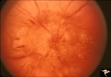
Chronic Papilledema with Pseudo Drusen | Left eye of 51 year old, 220 pound black woman. Pseudotumor cerebri, pseudo drusen, exudates. Anatomy: Optic disc. Pathology: Papilledema Disease/Diagnosis: Chronic papilledema with pseudo drusen | Image |
| 58 |
 |
Chronic Papilledema with Pseudo Drusen | Chronic papilledema with pseudo drusen. Residual choroidal folds. Pseudo drusen. Anatomy: Optic disc. Pathology: Papilledema. Disease/Diagnosis: Chronic papilledema with pseudo drusen. | Image |
| 59 |
 |
Chronic Papilledema with Pseudo Drusen | Left eye. Meningioma. Pseudo drusen from chronic papilledema. The patient's meningioma had blinded her left eye and caused chronic elevated intracranial pressure. Woman. Anatomy: Optic disc Pathology: Papilledema Disease/Diagnosis: Chronic papilledema with pseudo drusen | Image |
| 60 |
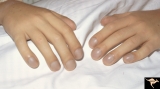 |
Cyanotic Heart Disease with Clubbing of Fingernails | Note the cyanotic nail beds and clubbing. Anatomy: Optic disc. Pathology: Papilledema. Disease/Diagnosis: Pseudotumor due to cyanotic heart disease. Clinical: Young boy with clubbing. | Image |
| 61 |
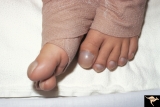 |
Cyanotic Heart Disease with Clubbing of Toes | Bilateral Papilledema with cyanotic heart disease. Anatomy: Optic disc. Pathology: Papilledema. Disease/Diagnosis: Pseudotumor due to cyanotic heart disease. Clinical: Young boy with clubbing. | Image |
| 62 |
 |
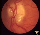
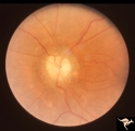
Medullated Nerve Fibers with Papilledema | Left eye. papilledema only. Man with metastatic gastric carcinoma. Anatomy: Optic disc. Pathology: Papilledema. Disease/Diagnosis: Papilledema plus medullated nerve fibers. | Image |
| 63 |
 |
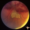
P50 Chronic Papilledema with Subretinal Neo-Vascular Network | Chronic papilledema with subretinal neo-vascular network. Pseudotumor. Anatomy: Optic disc. Pathology: Papilledema. Disease/ Diagnosis: Chronic papilledema with sub-retinal neovascular network. | Image |
| 64 |
 |
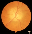
P52a Asymmetric Papilledema with Choroidal Folds | Left eye. Choroidal folds with no papilledema. Asymmetric papilledema with choroidal folds. Bilateral choroidal folds from elevated intracranial pressure. 52a. Anatomy: Optic disc. Pathology: Papilledema. Disease/ Diagnosis: Asymmetric - No papilledema with choroidal folds | Image |
| 65 |
 |
P52b Asymmetric Papilledema with Choroidal Folds | Right eye shows papilledema. Asymmetric papilledema with choroidal folds. Bilateral choroidal folds from elevated intracranial pressure. Anatomy: Optic disc. Pathology: Papilledema. Disease/ Diagnosis: Chronic papilledema with choroidal folds. | Image |
| 66 |
 |
P55a Chronic Papilledema, Late Stage | Right eye. Optic atrophy secondary to chronic papilledema. Glioblastoma. Chemotherapy patient. Anatomy: Optic disc. Pathology: Papilledema. Disease/ Diagnosis: Chronic papilledema with atrophy. | Image |
| 67 |
 |
P55b Chronic Papilledema, Late Stage | Left eye. Optic atrophy secondary to chronic papilledema, late stage with retinal choroidal bypass vein. Opticocilliary shunt. Glioblastoma, chemotherapy patient. Anatomy: Optic disc. Pathology: Papilledema. Disease/ Diagnosis: Chronic papilledema with atrophy. | Image |
| 68 |
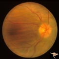 |
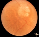
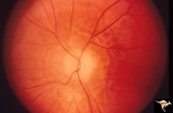
Post Papilledema with Choroidal Folds | Right eye. Post papilledema with choroidal folds due to brain tumor. Anatomy: Optic disc. Pathology: Post papilledema. Disease/Diagnosis: Post papilledema with choroidal folds. | Image |
| 69 |
 |
Resolution of Papilledema Following Optic Nerve Sheath Decompression (ONSD) | Left eye. 17 year old boy. Cryptococcal meningitis. Resolution of papilledema following optic nerve sheath decompression (ONSD) on November 1, 1974. Same eye as P_53a in January 1975. Atrophic, resolved disc. Note "high-water" marks. Visual acuity was 20/40. Anatomy: Optic disc. Pathology: Papilled... | Image |
| 70 |
 |
Resolution of Papilledema Following Optic Nerve Sheath Decompression (ONSD) | Left eye. 17 year old boy. Cryptococcal meningitis. Resolution of papilledema following optic nerve sheath decompression (ONSD) in November 1, 1974. Same eye as P_53a on December 1974. Atrophic. Note "high-water" marks. Anatomy: Optic disc. Pathology: Papilledema. Disease/Diagnosis: Resolving papill... | Image |
| 71 |
 |
Resolution of Papilledema Following Optic Nerve Sheath Decompression (ONSD) | Left eye. 17 year old boy. Cryptococcal meningitis. Module developed papilledema. June 1974. Anatomy: Optic disc. Pathology: Papilledema. Disease/Diagnosis: Resolving papilledema. | Image |
| 72 |
 |
Resolution of Papilledema Following Optic Nerve Sheath Decompression (ONSD) | Left eye. 17 year old boy. Cryptococcal meningitis. Resolution of papilledema following optic nerve sheath fenestration (ONSF) on November 1, 1974. Same eye as P_53a on November 7, 1974, one week following ONSF. Anatomy: Optic disc. Pathology: Papilledema. Disease/Diagnosis: Resolving papilledema. | Image |
| 73 |
 |
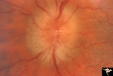
Resolution of Papilledema Following Optic Nerve Sheath Decompression (ONSD) | Left eye. 17 year old boy. Cryptococcal meningitis. Same eye as P_53a. Increased papilledema. August 1974. Anatomy: Optic disc. Pathology: Papilledema. Disease/Diagnosis: Resolving papilledema. | Image |
| 74 |
 |
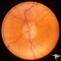
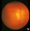
Unilateral Papilledema | Right eye. Has no optic cup. Disc is flat. Anatomy: Optic disc. Pathology: Unilateral papilledema. Disease/Diagnosis: Idiopathic intracranial hypertension (pseudotumor cerebri). Clinical: Transient monocular blindness (transient visual obscurations, amaurosis fugax); headache, sixth nerve palsy, ele... | Image |
| 75 |
 |
Unilateral Papilledema | Left eye. This eye has papilledema. Anatomy: Optic disc. Pathology: Unilateral papilledema. Disease/Diagnosis: Idiopathic intracranial hypertension (pseudotumor cerebri). Clinical: Transient monocular blindness (transient visual obscurations, amaurosis fugax); headache, sixth nerve palsy, elevated i... | Image |