Best known for his world-renowned neuro-ophthalmology unit based at the University of California, San Francisco, William Hoyt, MD collected here more than 850 of his best images covering a wide range of disorders.
William F. Hoyt, MD, Professor Emeritus of Ophthalmology, Neurology and Neurosurgery, Department of Ophthalmology, University of California, San Francisco.
NOVEL: https://novel.utah.edu/
TO
| Title | Description | Type | ||
|---|---|---|---|---|
| 576 |
 |
Venous Anomalies - Exit Anomalies | Choriovaginal vein. Choroid in myopic disc is draining into optic disc. Choriovaginal vein entering disc ege at 9:00. Kraupa type 2.Peripapillary atrophy in highly myopic eye. Same patient as V_39. Anatomy: Optic disc. Pathology: Congenital anomaly of choroidal venous drainage. Disease/Diagnosis: C... | Image |
| 577 |
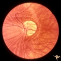 |
Venous Anomalies - Exit Anomalies | Vein crosses entire disc to disc edge, possibly into choroidal vein at 3:30 Disc edge veins of Kraupa. Anatomy: Optic disc. Pathology: Congenital anomaly, exit anomaly. Disease/Diagnosis: Exit anomaly, edge veins. Clinical: Asymptomatic. | Image |
| 578 |
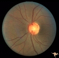 |
Venous Anomalies - Exit Anomalies | Disc edge veins of Kraupa. 35 year old woman. Note that the arterial branches all appear to be cilioretinal. Empty disc. Anatomy: Optic disc. Pathology: Congenital anomaly, exit anomaly. Disease/Diagnosis: Exit anomaly, edge veins. Clinical: Asymptomatic. | Image |
| 579 |
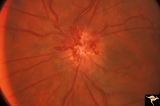 |
Venous Anomalies - Prepapillary Venous Convolutions (Acquired) | Prepapillary venous convolutions - acquired. Acquired after central retinal vein occlusion. Anatomy: Optic disc. Pathology: Prepapillary venous convolutions - acquired. Disease/Diagnosis: Prepapillary venous convolutions - acquired. Clinical: Asymptomatic. | Image |
| 580 |
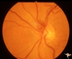 |
Venous Anomalies - Prepapillary Venous Convolutions (Congenital) | Sub-retinal and prepapillary venous convolutions - congenital. Edge vein. Large vein draining subretinally into the choroid. Anatomy: Optic disc. Pathology: Prepapillary venous convolutions - congenital. Disease/Diagnosis: Prepapillary venous convolutions - congenital. Clinical: Asymptomatic. | Image |
| 581 |
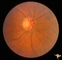 |
Venous Anomalies - Prepapillary Venous Convolutions (Congenital) | Prepapillary venous convolutions - congenital. Anatomy: Optic disc. Pathology: Prepapillary venous convolutions - congenital. Disease/Diagnosis: Prepapillary venous convolutions - congenital. Clinical: Asymptomatic. | Image |
| 582 |
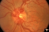 |
Venous Anomalies - Prepapillary Venous Convolutions (Congenital) | Prepapillary venous convolutions - congenital. 42 year old man. Incidental finding. Anatomy: Optic disc. Pathology: Prepapillary venous convolutions - congenital. Disease/Diagnosis: Prepapillary venous convolutions - congenital. Clinical: Asymptomatic. | Image |
| 583 |
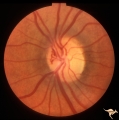 |
Venous Anomalies - Prepapillary Venous Convolutions (Congenital) | Prepapillary venous convolutions - congenital. Anatomy: Optic disc. Pathology: Prepapillary venous convolutions - congenital. Disease/Diagnosis: Prepapillary venous convolutions - congenital. Clinical: Asymptomatic. | Image |
| 584 |
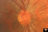 |
Venous Anomalies - Prepapillary Venous Convolutions (Congenital) | Prepapillary venous convolutions - congenital. Anatomy: Optic disc. Pathology: Prepapillary venous convolutions - congenital. Disease/Diagnosis: Prepapillary venous convolutions - congenital. Clinical: Asymptomatic. | Image |
| 585 |
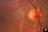 |
Venous Anomalies - Prepapillary Venous Convolutions (Congenital) | Prepapillary venous loop - congenital. Anatomy: Optic disc. Pathology: Prepapillary venous convolutions - congenital. Disease/Diagnosis: Prepapillary venous convolutions - congenital. Clinical: Asymptomatic. | Image |
| 586 |
 |
Visible Drusen | PP21a: Right eye. Drusen barely visible. Note disc margin drusen at 1:00 and 2:30.; PP21b: Left eye shows multiple exposed drusen. Girl. Anatomy: Optic disc. Pathology: Drusen of the optic disc. Disease/Diagnosis: Drusen of the optic disc. Clinical: Normally functioning eye with drusen. | Image |
| 587 |
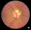 |
Visible Drusen | PP24a. Right eye. Exposed drusen. There are inferior nerve fiber layer defects in the upper arcuate bundles. Optic disc is also hypoplastic. Anatomy: Optic disc. Pathology: Drusen of the optic disc. Disease/Diagnosis: Drusen of the optic disc. Clinical: Hypoplastic optic disc with drusen. | Image |
| 588 |
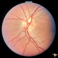 |
Visible Drusen - Bilateral | PP22a: right eye. PP22b: Note bypass vein draining into the choroid at 8:00. Anatomy: Optic disc. Pathology: Drusen of the optic disc. Disease/Diagnosis: Drusen of the optic disc. Clinical: Normally functioning eye with drusen. | Image |
| 589 |
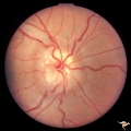 |
Visible Drusen - Bilateral | PP22a: right eye with obvious exposed drusen. PP22 b: Note bypass vein draining into the choroid at 8:00. Anatomy: Optic disc. Pathology: Drusen of the optic disc. Disease/Diagnosis: Drusen of the optic disc. Clinical: Normally functioning eye with drusen. | Image |
| 590 |
 |
Visible Drusen with Visual Field Loss | Right eye visual field combine with PP25a, b, & d. Anatomy: Optic disc. Pathology: Drusen of the optic disc. Disease/Diagnosis: Drusen of the optic disc. Clinical: Drusen disc with severe visual field defect. note the nasal visual field loss and the arcuate bundle defects. Central vision was 20/20. | Image |
| 591 |
 |
Visible Drusen with Visual Field Loss | Left eye visual field. Combine with PP25 a, b, & c. Anatomy: Optic disc. Pathology: Drusen of the optic disc. Disease/Diagnosis: Drusen of the optic disc. Clinical: Note marked constriction of visual field in all quadrants with central preservation of vision. | Image |
| 592 |
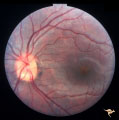 |
Visible Drusen with Visual Field Loss | Left eye.16 year old girl: PP26b: buried drusen at the lower pole of the disc; PP26a: Visible drusen with visual field loss. Notice the thinning of the nerve fibers in both the superior and inferior arcuate bundles. PP26c: Goldmann visual field. Anatomy: Optic disc. Pathology: Drusen of the optic ... | Image |
| 593 |
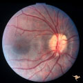 |
Visible Drusen with Visual Field Loss | Right eye.16 year old girl: PP26a: Visible drusen with visual field loss. Notice the thinning of the nerve fibers in both the superior and inferior arcuate bundles. PP26b: buried drusen; PP26c: Goldmann visual field. Anatomy: Optic disc. Pathology: Drusen of the optic disc. Disease/Diagnosis: Druse... | Image |
| 594 |
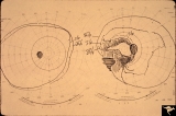 |
Visible Drusen with Visual Field Loss | 16 year old girl: Drusen disc. Goldmann visual field. Anatomy: Optic disc. Pathology: Drusen of the optic disc. Disease/Diagnosis: Drusen of the optic disc. Clinical: Drusen disc with visual field loss. | Image |
| 595 |
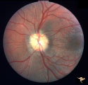 |
Visible Drusen with Visual Field Loss | PP25a: Left eye: Severe visual field defect. PP25b: right eye with exposed drusen and field loss: visual field defects; PP25c: right eye visual field PP25d: left eye visual field. Anatomy: Optic disc. Pathology: Drusen of the optic disc. Disease/Diagnosis: Drusen of the optic disc. Clinical: Dr... | Image |
| 596 |
 |
Visible Drusen with Visual Field Loss | PP25b right eye with drusen and severe visual field loss. Match with PP25a, c & d. Anatomy: Optic disc. Pathology: Drusen of the optic disc. Disease/Diagnosis: Drusen of the optic disc. Clinical: Drusen disc with servere visual field loss. | Image |
| 597 |
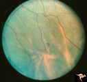 |
Von Hippel Lindau Disease | Von Hippel Lindau Disease with a mini retinal tumor. Pair with R1_C4b. Anatomy: Retina. Pathology: Hemangioblastoma. Disease/Diagnosis: Von Hippel Lindau disease. Clinical: No visual symptoms. Imaging: Flourescien angiogram in R1_C4b. | Image |
| 598 |
 |
Von Hippel Lindau Disease | Von Hippel Lindau lesion on optic disc showing minimal increase in size over three year interval. Anatomy: Optic disc. Pathology: Hemangioblastoma. Disease/Diagnosis: Von Hippel Lindau disease. Clinical: Patient other eye was removed for hemangioblastoma. He has numerous hemangioblastomas of his spi... | Image |
| 599 |
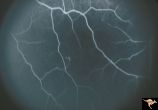 |
Von Hippel Lindau Disease | Von Hippel Lindau Disease with a mini retinal tumor. Flourescien angiogram shows how small tumor is. Pair with R1_C4a. Anatomy: Retina. Pathology: Hemangioblastoma. Disease/Diagnosis: Von Hippel Lindau disease. Clinical: No visual symptoms. Imaging: Flourescien angiogram. | Image |
| 600 |
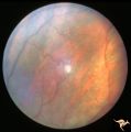 |
Von Hippel Lindau Disease | Von Hippel Lindau Disease. Retinal photograph showing small whitish hemangiomablastoma. Note the dilated arterial and venous channels entering and leaving the tumor. Anatomy: Retina. Pathology: Hemangioblastoma. Disease/Diagnosis: Von Hippel Lindau disease. Clinical: No visual symptoms. | Image |
