Best known for his world-renowned neuro-ophthalmology unit based at the University of California, San Francisco, William Hoyt, MD collected here more than 850 of his best images covering a wide range of disorders.
William F. Hoyt, MD, Professor Emeritus of Ophthalmology, Neurology and Neurosurgery, Department of Ophthalmology, University of California, San Francisco.
NOVEL: https://novel.utah.edu/
TO
Filters: Collection: "ehsl_novel_wfh"
| Title | Description | Type | ||
|---|---|---|---|---|
| 101 |
 |
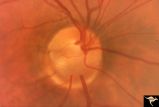
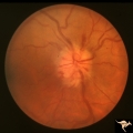
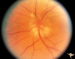
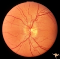
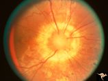
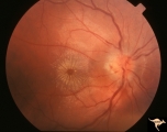
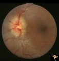
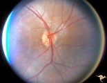
Buried Drusen | Young woman with pseudo papilledema from buried drusen with associated visual field defects. Barely visible in the upper arcuate nerve fibers is a slit like defect. Anatomy: Optic disc. Pathology: Drusen of the optic disc. Disease/Diagnosis: Drusen of the optic disc. Clinical notes: This patient had... | Image |
| 102 |
 |
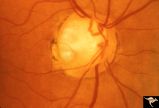
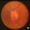
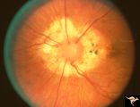
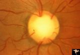
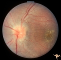
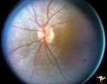
Buried Drusen | Buried drusen with peculiar white dot, which appears to be choroidal in location. Note lumpy disc margin on right disc PP_15a is right eye. PP_15b is left eye. Beautiful example of pseudo papilledema in which drusen can not be seen. 8 year old girl. Anatomy: Optic disc. Pathology: Drusen of the op... | Image |
| 103 |
 |
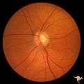
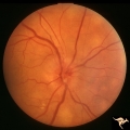
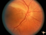
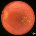
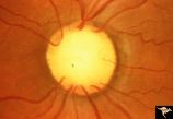
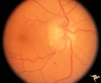
Buried Drusen | Suspected buried drusen in a girl. Anatomy: Optic disc. Pathology: Drusen of the optic disc. Disease/Diagnosis: Drusen of the optic disc. Clinical notes: Normally functioning eye with suspected drusen. | Image |
| 104 |
 |
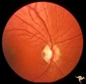
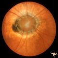
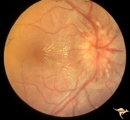
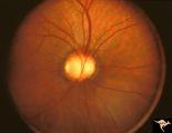
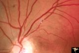
Buried Drusen | Left disc has a blurred lumpy margin. Retinal vessels are not obscured in the disc margin blur, therefore no edema is present. This is an example of a difficult blurred disc, the nature of which is clarified by the presence of a clear cut disk anomoly in the fellow eye. 8 year old girl. PP_15a has b... | Image |
| 105 |
 |
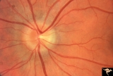
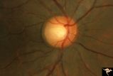
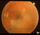
Buried Drusen | Buried drusen; PP_13a: Right eye. Note lumpy disc margin, especially temporally. Also note absence of optic cup. Excellent example of pseudo papilledema with buried drusen. Anatomy: Optic disc. Pathology: Drusen of the optic disc. Disease/Diagnosis: Drusen of the optic disc. Clinical notes: Patient ... | Image |
| 106 |
 |
Buried Drusen | Buried drusen. Left eye. Note lumpy disc margin, especially temporally. Also note absence of optic cup. Excellent example of pseudo papilledema with buried drusen. Pair with PP_13a. Anatomy: Optic disc. Pathology: Drusen of the optic disc. Disease/Diagnosis: Drusen of the optic disc. Clinical notes... | Image |
| 107 |
 |
Buried Drusen | Excellent example of pseudo papilledema with sub surface drusen at 10:00 and 1:00. Anatomy: Optic disc. Pathology: Drusen of the optic disc. Disease/Diagnosis: Drusen of the optic disc. Clinical notes: Normally functioning eye with drusen. | Image |
| 108 |
 |
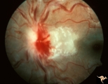
Buried Drusen with Choroidal Retinal Scar | Right eye: Buried drusen; probable complication of peripapillary hemorrhage at 7:00. Anatomy: Optic disc. Pathology: Drusen of the optic disc. Disease/Diagnosis: Drusen of the optic disc. Clinical notes: Enlarged blind spot. | Image |
| 109 |
 |
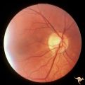
Buried Drusen with Sub-retinal Neovascular Net | Buried drusen with sub-retinal neovascular net. Both PP29a and PP29b are left eye: 17 year old girl; Visual acuity 10/400. Anatomy: Optic disc. Pathology: Drusen of the optic disc. Disease/Diagnosis: Drusen of the optic disc. Clinical notes: Loss of central vision due to subretinal neovascularizatio... | Image |
| 110 |
 |
Buried Drusen with Sub-retinal Neovascular Net | Buried drusen with sub-retinal neovascular net. This is the same left eye. Appearance of the central retina of the left eye. Both PP29a & b are left eye: 17 year old girl; Visual acuity 10/400. Anatomy: Optic disc. Pathology: Drusen of the optic disc. DIsease/Diagnosis: Drusen of the optic disc. Cl... | Image |
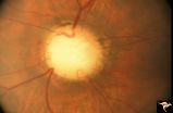
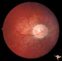
| 111 |
 |
C01 Pits of the Optic Disc | Right eye. Very large inferior temporal optic pit. Congenital. Woman. Anatomy: Optic disc. | Image |
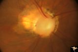
| 112 |
 |
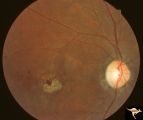
C02 Pits of the Optic Disc | Right eye. Three congenital optic pits on the temporal side. 8:00, 9:30, 10:30. Anatomy: Optic disc. | Image |
| 113 |
 |
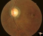
C03 Pits of the Optic Disc | Central optic pit. Left eye. Anatomy: Optic disc. | Image |
| 114 |
 |
C04 Pits of the Optic Disc | Right eye. Man. Large temporal pit. Macular detachment. Anatomy: Optic disc. | Image |
| 115 |
 |
C05 Pits of the Optic Disc | Right eye. Pigmented pit. Woman. Anatomy: Optic disc. | Image |
| 116 |
 |
C06 Pits of the Optic Disc | Right eye. Temporal pit. 6 year old with see-saw nystagmus. Anatomy: Optic disc. Clinical: Six-year old with see-saw nystagmus. | Image |
| 117 |
 |
C07 Pits of the Optic Disc | Left eye. Temporal pit. Man. Anatomy: Optic disc. | Image |
| 118 |
 |
C08 Pits of the Optic Disc | Left eye. Large cavitary anomaly (pit). Man. 20/100 visual acuity. Superior nasal visual field defect. May not have a central retinal artery. Anatomy: Optic disc. Clinical: Man. 20/100 visual acuity. Superior nasal visual field defect. | Image |
| 119 |
 |
C09 Pits of the Optic Disc | Pit with peripapillary choroidal defect. Right eye. Dwarfed boy. May not have a central retinal artery. Same patient as C_10. Anatomy: Optic disc. | Image |
| 120 |
 |
C10 Pits of the Optic Disc | Disc malformation. Abortive cavitary anomaly. Left eye. Dwarfed boy. Same patient as C_9. Anatomy: Optic disc. | Image |
| 121 |
 |
C101 Papillitis, Retrobulbar Neuritis | Resolved. Associated polycythemia. Papillitis after flu in patient with polycythemia. Homosexual male. Anatomy: Optic disc. Pathology: Axoplasmic stasis due to inflammation. Disease/ Diagnosis: Post infectious papillitis. Clinical: Visual loss after the flu.. | Image |
| 122 |
 |
C102 Papillitis, Retrobulbar Neuritis | Inflammatory papillitis in 25 year old woman. Resolved completely. Anatomy: Optic disc. Pathology: Axoplasmic stasis due to inflammation. Disease/ Diagnosis: Inflammatory optic papillitis. Clinical: Visual loss. | Image |
| 123 |
 |
C103 Papillitis, Retrobulbar Neuritis | Optic neuritis in infectious mononucleosis. Anatomy: Optic disc. Pathology: Axoplasmic stasis due to inflammation. Disease/ Diagnosis: Optic neuritis with mononucleosis or Epstein Barr Virus. Clinical: Visual loss associated with mononucleosis. | Image |
| 124 |
 |
C104 Papillitis, Retrobulbar Neuritis | Post infectious papillitis with macular exudate. Anatomy: Optic disc macula. Pathology: Axoplasmic stasis due to inflammation with lipid deposit in Henle's layer. Disease/ Diagnosis: Post infectious papillitis/optic neuritis. Clinical: Visual loss after infection. | Image |
| 125 |
 |
C105 Disc Edema with Systemic Lupus | Mild disc edema blurs the inferior disc margin. Flourescein angiogram in D1_06. Same patient as D1_06 an D1_07. Man. Anatomy: Optic disc. Pathology: Axoplasmic stasis due to vasculitis (Lupus). Disease/ Diagnosis: Lupus papillopathy. | Image |
| 126 |
 |
C106 Papillitis, Retrobulbar Neuritis | Papillitis with recovery of vision. Woman acupuncturist. Anatomy: Optic disc. Pathology: Axoplasmic stasis after inflammation. Disease/ Diagnosis: Optic neuritis/optic papillitis. Clinical: Visual loss with recovery. | Image |
| 127 |
 |
C107 Papillitis, Retrobulbar Neuritis | Man with bilateral papillitis. Right eye. Pair with C1_08. Cause unknown. Visual field showed central scotomas. Anatomy: Optic disc. Pathology: Axoplasmic stasis due to inflammation. Disease/ Diagnosis: Neuritis of the optic nerve. Clinical: Visual loss. | Image |
| 128 |
 |
C108 Papillitis, Retrobulbar Neuritis | Man with bilateral papillitis. Left eye. Pair with C1_07. Cause unknown. Visual field shows central scotoma. Anatomy: Optic disc. Pathology: Axoplasmic stasis due to inflammation. Disease/ Diagnosis: Optic neuritis / Optic papillitis. Clinical: Visual loss. | Image |
| 129 |
 |
C109 Papillitis, Retrobulbar Neuritis | Optic papillitis after wasp sting. 57 year old woman. Right eye. Anatomy: Optic disc. Pathology: Axoplasmic stasis due to inflammation. Disease/ Diagnosis: Optic neuritis after wasp sting. Clinical: Visual loss after wasp sting. | Image |
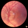
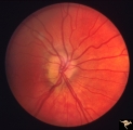
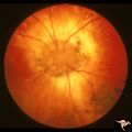
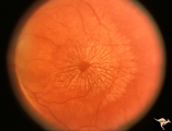
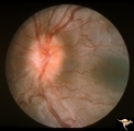
| 130 |
 |
C11 Morning Glory Disc | "Morning Glory" disc. 11 year old girl. May not have a central retinal artery. Anatomy: Optic disc. | Image |
| 131 |
 |
C110 Papillitis, Retrobulbar Neuritis | AIDs papillitis. Segmental. Note inflammatory focus on temporal side of disc. 29 year old homosexual male. Visual field shows huge blind spot. Anatomy: Optic disc. Pathology: Axoplasmic stasis due to inflammation. Disease/ Diagnosis: AIDS papillitis / AIDS Optic neuritis. Clinical: Visual symptoms d... | Image |
| 132 |
 |
C111 Papillitis, Retrobulbar Neuritis | AIDS papillitis. Male. Anatomy: Optic disc. Pathology: Axoplasmic stasis due to inflammation. Disease/ Diagnosis: AIDS papillitis. Clinical: Visual symptoms. | Image |
| 133 |
 |
C112 Papillitis, Retrobulbar Neuritis | Woman with herpes. Acute retinal necrosis with papillitis and arcuate neuro-retinitis. Right eye. Pair with C1_13. Reference: Margolis T, Irvine AR, Hoyt WF, Hyman R. Acute retinal necrosis syndrome presenting with papillitis and arcuate neuroretinitis. Ophthalmology. 1988 Jul;95(7):937-40. Anatomy:... | Image |
| 134 |
 |
C113 Papillitis, Retrobulbar Neuritis | Woman with herpes. Acute retinal necrosis with papillitis an arcuate neuro-retinitis. Right eye. Notice the large arcuate defect extending fromt he disc to the retina of retinal necrosis. Pair with C1_12. Anatomy: Optic disc; Retina. Pathology: Axoplasmic stasis due to inflammation; Retinal necrosis... | Image |
| 135 |
 |
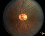
C114 Papillitis, Retrobulbar Neuritis | Epstien-Barr Virus papillitis with remarkably good recovery. Steroid responsive. Woman. Anatomy: Optic disc. Pathology: Axoplasmic stasis due to inflammation. Disease/ Diagnosis: Epstein Barr Virus with papillitis. Clinical: Visual loss that was steroid responsive. | Image |
| 136 |
 |
C115 Papillitis, Retrobulbar Neuritis | Demyelinative optic neuropathy with mild disc swelling. This eye had a large central scotoma. Note the bland disc margin swelling from 2:00 to 4:00. This swelling constitutes spill over edema from the main focus of the neuritis which lies behind the eyeball. Visual acuity was 2200. Anatomy: Optic di... | Image |
| 137 |
 |
C116 Papillitis, Retrobulbar Neuritis | Demyelinative optic neuropathy with mild disc swelling. Note the mild disc margin blurring. The main focus of the neuritis lies behind the eye. 20/20 visual acuity. Anatomy: Optic disc; Optic nerve. Pathology: Axoplasmic stasis due to inflammation; Acute demyelination. Disease/ Diagnosis: Acute sub-... | Image |
| 138 |
 |
C12 Morning Glory Disc | "Morning Glory" disc. Patient 11 year old. Anatomy: Optic disc. | Image |
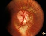
| 139 |
 |
C13 Morning Glory Disc | "Morning Glory" disc with peripapillary choroidal defect extending inferiorly. Patient has transphenoidal encephalocele. Note tapering edge like an arrow pointing to patient's basal encephalocele and cleft palate. Reference: Brodsky MC, Hoyt WF, Hoyt CS, Miller NR, Lam BL. Atypical retinochoroidal ... | Image |
| 140 |
 |
C14 Morning Glory Disc | Isolated "Morning Glory". Left eye. Girl. Anatomy: Optic disc. | Image |
| 141 |
 |
C15 Morning Glory Disc | "Morning Glory" disc. Note tapering edge pointing to patient's transphenoidal encephalocele. Reference: Brodsky MC, Hoyt WF, Hoyt CS, Miller NR, Lam BL. Atypical retinochoroidal coloboma in patients with dysplastic optic discs and transphenoidal encephalocele Arch Ophthalmol. 1995 May;113(5):624-8.... | Image |
| 142 |
 |
C16 Morning Glory Disc | "Morning Glory" disc. Note tapering edge pointing to basal encephalocele. Boy. Anatomy: Optic disc. | Image |
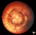
| 143 |
 |
C17 Morning Glory Disc | "Morning Glory" disc. CT normal. Anatomy: Optic disc. Clinical: CT normal. | Image |
| 144 |
 |
C18 Morning Glory Disc | "Morning Glory" disc. 6 month old baby. Anatomy: Optic disc | Image |
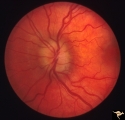
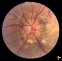
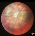
| 145 |
 |
C19 Morning Glory Disc | Bilateral "Morning Glory" disc. Right eye. Man. Pair with C_20. Anatomy: Optic disc. | Image |
| 146 |
 |
C20 Morning Glory Disc | Bilateral "Morning Glory" disc. Left eye. Man. Pair with C_19. Anatomy: Optic disc. | Image |
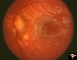
| 147 |
 |
C201 Papillitis with Macular Star, Cat Scratch Disease | Proven Bartonella neuroretinitis. Notice the deposit of exudates of Henle's layer making an almost complete macular star. Anatomy: Optic disc; Retina. Pathology: Neuroretinitis; Axoplasmic stasis due to inflammation. Disease/ Diagnosis: Neuroretinitis due to Bartonella Henslae. Clinical: Visual blur... | Image |
| 148 |
 |
C202 Papillitis with Macular Star Cat Scratch Disease. | Proven Bartonella neuroretinitis. 23 year old man. Ocular disc edema with macular star (ODEMS). Anatomy: Optic disc; Retina. Pathology: Axoplasmic stasis due to inflammation; Exudate in Henle's layer. Neuroretinitis due to Bartonella Henslae (or cat scratch). Clinical: Visual blurring; Optic disc sw... | Image |
| 149 |
 |
C203 Papillitis with Macular Star, Cat Scratch Disease | Proven Bartonella neuroretinitis. Left eye. October 3, 1986. Same eye as C2_04. Macular star visible on C2_04. Woman. Ocular disc edema with macular star (ODEMS). Anatomy: Optic disc; Retina. Pathology: Axoplasmic stasis due to inflammation; Exudates in Henle's layer. Disease/ Diagnosis: Bartonella ... | Image |
| 150 |
 |
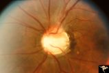
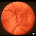
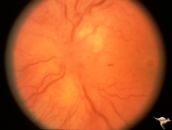
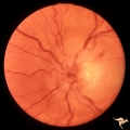
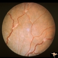
C204 Papillitis with Macular Star, Cat Scratch Disease | Proven Bartonella neuroretinitis. Left eye. October 17, 1986. Same eye as C2_03. Ocular disc edema with macular star (ODEMS). Woman. Anatomy: Optic disc; Retina. Pathology: Exudates in Henle's layer. DIsease/ Diagnosis: Neuroretinitis due to Bartonella Henslae (Cat Scratch). Clinical: Visual blurrin... | Image |
| 151 |
 |
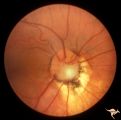
C205 Papillitis with Macular Star, Cat Scratch Disease | Proven Bartonella neuroretinitis. Macular star present, but not visible on image. 33 year old woman. Anatomy: Optic disc; Retina. Pathology: Axoplasmic stasis due to inflammation. Disease/ Diagnosis: Bartonella Henslae (Cat Scratch). Clinical: Visual blurring. | Image |
| 152 |
 |
C206 Papillitis with Macular Star, Cat Scratch Disease | Proven Bartonella neuroretinitis. Resolved papillitis with residual retinal exudate. Man. Anatomy: Optic disc; Retina. Pathology: Axoplasmic stasis due to inflammation. Disease/ Diagnosis: Bartonella Henslae (Cat Scratch). Clinical: Visual blurring; Ocular edema with macular star (ODEMS). | Image |
| 153 |
 |
C207 Papillitis with Macular Star, Cat Scratch Disease | Proven Bartonella neuroretinitis. Anatomy: Optic disc; Retina. Pathology: Axoplasmic stasis due to inflammation. Disease/ Diagnosis: Bilateral Bartonella Henslae (Cat Scratch). Clinical: Visual blurring; Ocular disc edema with macular star (ODEMS). | Image |
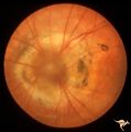
| 154 |
 |
C208 Papillitis with Macular Star, Cat Scratch Disease | Proven Bartonella neuroretinitis. Man. Anatomy: Optic disc; Retinitis. Pathology: Axoplasmic stasis due to inflammation. Disease/ Diagnosis: Bartonella Henslae (Cat Scratch). Clinical: Visual blurring; Ocular disc edema with macular star (ODEMS). | Image |
| 155 |
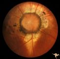
 |
C209 Papillitis with Macular Star, Cat Scratch Disease | Proven Bartonella neuroretinitis. Woman. Anatomy: Optic disc; Retina. Pathology: Axoplasmic stasis due to inflammation. Disease/ Diagnosis: Bartonella Henslae (Cat Scratch). Clinical: Visual blurring without visual field defect; Ocular disc edema with macular star (ODEMS). | Image |
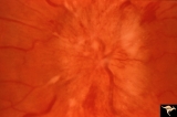
| 156 |
 |
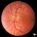
C21 Empty Disc | Right eye. All cilioretinal fundus. No central retinal artery. Handmann anomaly. Frequently associated with renal dysplasia. Pair with C_22 an C_23. Reference: Barroso LH, Hoyt WF, Narahara M. Can the arterial supply of the retina in man be exclusively cilioretinal? J Neuroophthalmol. 1994 Jun;14(2... | Image |
| 157 |
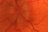
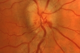
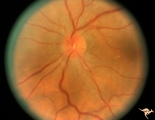
 |
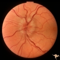
C22 Empty Disc | Left eye. All cilioretinal fundus. No central retinal artery. Handmann anomaly. Frequently associated with renal dysplasia. Pair with C_21 an C_23. Reference: Barroso LH, Hoyt WF, Narahara M. Can the arterial supply of the retina in man be exclusively cilioretinal? J Neuroophthalmol. 1994 Jun;14(2)... | Image |
| 158 |
 |
C23 Empty Disc | Father of patient in C_21 and C_22. Father has central retinal artery, multiple cilioretinal arteries and had previously unsuspected renal failure. Papillorenal Syndrome (PRS). Reference: Parsa,CF et al. Ophthalmology. 2001. 108(4): 738-49Barroso LH, Hoyt WF, Narahara M. Can the arterial supply of ... | Image |
| 159 |
 |
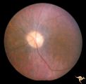
C24 Empty Disc | Left eye. Multiple cilioretinal arteries. Child of C_25 Anatomy: Optic disc. | Image |
| 160 |
 |
C25 Empty Disc | Right eye. Multiple cilioretinal arteries. Visual function normal. Father of C_24. Anatomy: Optic disc | Image |
| 161 |
 |
C26 Empty Disc | Right eye. Multiple cilioretinal arteries. Patient had dysplastic kidneys. Papillorenal Syndrome (PRS). Hand motion vision. 17 year old girl. Anatomy: Optic disc. | Image |
| 162 |
 |
C27 Empty Disc | Multiple cilioretinal arteries. Chronic interstitial nephritis. Renal and optic disc dysplasia. Papilorenal Syndrome (PRS). No central retinal artery. Anatomy: Optic disc. | Image |
| 163 |
 |
C28 Empty Disc | Left eye. Woman. Multiple cilioretinal vessels. Visual function normal. Anatomy: Optic disc. | Image |
| 164 |
 |
C29 Empty Disc | Left eye. Papilorenal Syndrome (PRS). Pair with C_30. Anatomy: Optic disc. | Image |
| 165 |
 |
C30 Empty Disc | Right eye. Papillorenal Syndrome (PRS). Pair with C_29. Anatomy: Optic disc. | Image |
| 166 |
 |
C301 Nodular Papillopathies (Sarcoid) | Disc swelling. Sarcoid papillopathy. Note infiltrative nodule at 9:00 on the disc.The patient had proven sarcoid. Perivenous inflammatory cuffing visible on image C3_02. Right eye. Pair with C3_02. Anatomy: Optic disc; Retina. Pathology: Axoplasmic stasis due to sarcoid infiltration. Disease/ Diagn... | Image |
| 167 |
 |
C302 Nodular Papillopathies (Sarcoid) | Perivenous Inflammatory Cuffing in a Patient with Proven Sarcoid. Left eye. Pair with C3_01. Anatomy: Retina. Pathology: Axoplasmic stasis due to sarcoid infiltration with retinal venous exudation? Disease/ Diagnosis: Sarcoid papillopathy with perivenous inflammatory disease. Clinical: Unknown? | Image |
| 168 |
 |
C303 Nodular Papillopathies (Sarcoid) | Lumpy infiltrative papillopathy in a patient with proven sarcoid. Anatomy: Optic disc. Pathology: Axoplasmic stasis due to sarcoid infiltration. Disease/ Diagnosis: Sarcoid papillopathy. Clinical: Unknown? | Image |
| 169 |
 |
C304 Nodular Papillopathies (Sarcoid) | Lumpy nodular disc infiltration from sarcoid. Anatomy: Optic disc. Pathology: Axoplasmic stasis due to sarcoid infiltration. Disease/ Diagnosis: Sarcoid papillopathy. Clinical: Unknown? | Image |
| 170 |
 |
C305 Nodular Papillopathies (Sarcoid) | July 1984 shows multiple infiltrative nodules on the optic disc in addition to circumferential subretinal yellow exudates. 32 year old black woman. Same patient as C3_06 and C3_07. Anatomy: Optic disc; Retina. Pathology: Axoplasmic stasis due to sarcoid infiltration and retinal exudation. Disease/ ... | Image |
| 171 |
 |
C306 Nodular Papillopathies (Sarcoid) | Lumpy disc swelling with retinal folds and a macular star in a patient with sarcoid. Presentation in October 1983. Same patient as C3_05 and C3_07. Anatomy: Optic disc; Retina. Pathology: Axoplasmic stasis due to sarcoid infiltration. Disease/ Diagnosis: Axoplasmic stasis due to sarcoid infiltration... | Image |
| 172 |
 |
C307 Nodular Papillopathies (Sarcoid) | Fluorescein angiogram shows striking staining of the sarcoid nodules. July 1984. Same patient as C3_05 and C3_06. Corresponds with July 1984 image, C3_06. Anatomy: Optic disc. Pathology: Axoplasmic stasis due to sarcoid infiltration. Disease/ Diagnosis: Sarcoid papillopathy. Clinical: Unknown? | Image |
| 173 |
 |
C308 Nodular Papillopathies (Sarcoid) | Nodular infiltrative papillopathy in a patient with sarcoid. Woman. Anatomy: Optic disc. Pathology: Axoplasmic stasis due to sarcoid infiltration. Disease/ Diagnosis: Sarcoid papillopahty. Clinical: Unknown? | Image |
| 174 |
 |
C31 Empty Disc | Right eye. Papillorenal Syndrome (PRS). Same patient as C_32. Anatomy: Optic disc. | Image |
| 175 |
 |
C32 Empty Disc | Left eye. Papillorenal Syndrome (PRS). Same patient as C_31. Anatomy: Optic disc. | Image |
| 176 |
 |
C33 Anomalous Pale Disc | Woman. Multiple cilioretinal arteries. Veins all empty into eye. Anomalous venous exit from nasal edge of optic disc. Visual function normal. Pair with C_36. Anatomy: Optic disc. | Image |
| 177 |
 |
C34 Anomalous Pale Disc | Multiple cilioretinal arteries. Anomalous venous exit from nasal edge of optic disc (Vein of Kraupa). Visual function normal. Anatomy: Optic disc. | Image |
| 178 |
 |
C35 Anomalous Pale Disc | Macro disc appears pale because of large diameter. Woman. Right eye. Anatomy: Optic disc. | Image |
| 179 |
 |
C36 Anomalous Pale Disc | Multiple cilioretinal arteries. Pale appearance. Normal optic nerve function. Good example of "empty disc". Pair with C_33. Anatomy: Optic disc. | Image |
| 180 |
 |
C37 Anomalous Pale Disc | "Watermelon" disc. Woman. Normal function. Left eye. Anatomy: Optic disc. | Image |
| 181 |
 |
C38 Anomalous Pale Disc | Megalopapilla in -8 myopic eye. Right eye. Anatomy: Optic disc. Clinical: High myope. | Image |
| 182 |
 |
C401 Luetic Papillopathy (Gumma of the Optic Disc) | Diffuse optic disc swelling with tortuous capillary dilations indicating inflammatory cellular infiltration. October 2001. Same eye as C4_02. Anatomy: Optic disc. Pathology: Axoplasmic stasis due to syphillitic infection. Luetic papillopathy (Syphyllis). Clinical: Visual loss. | Image |
| 183 |
 |
C402 Luetic Papillopathy (Gumma of the Optic Disc) | November 2001. Same eye as C4_01 after treatment with penicillin. Disc swelling went away and good visual function returned. Anatomy: Optic disc. Pathology: Axoplasmic stasis due to syphillitic infection. Disease/ Diagnosis: Luetic papillopathy (Syphillis). Clinical: Improving visual loss. | Image |
| 184 |
 |
C403 Luetic Papillopathy (Gumma of the Optic Disc) | 40 year old man with AIDS and neurosyphillis with severe visual field defect. The disc is pale and swollen and its arteries are strikingly narrowed (syphillitic vasculitis). Anatomy: Optic disc. Pathology: Axoplasmic stasis due to syphillitic infection. Disease/ Diagnosis: Luetic papillopathy (Syphy... | Image |
| 185 |
 |
Cerebellar Macular Degeneration | Cerebellar retinal degeneration with narrowed arterioles. Disc pallor. Granular retinal degeneration. 10 year old boy with mental degenerations and seizures. Anatomy: Retina. Pathology: Optic atrophy. Disease/Diagnosis: Congenital retinal cerebellar degeneration. Clinical: Severe mental retardation ... | Image |
| 186 |
 |
Cerebellar Macular Degeneration | Cerebellar retinal degenerative disease in a 12 year old boy who was blind and demented. His siblings were also blind. Was referred to as Voght-Spielmeyer Disease (Pair with R2_B1_3b shows granular retinal degeneration.) Anatomy: Retina. Pathology: Optic atrophy. Disease/Diagnosis: Congenital retina... | Image |
| 187 |
 |
Cerebellar Macular Degeneration | Cerebellar macular degeneration in a 7 year old boy with blindness. Rectal biopsy positive for storage material. Nature of cerebral degeneration was not defined in era when picture was taken. Sister also had similar findings. Anatomy: Retina. Pathology: Optic atrophy. Disease/Diagnosis: Congenital r... | Image |
| 188 |
 |
Cerebellar Macular Degeneration | Cerebellar retinal degenerative disease in a 12 year old boy who was blind and demented. His siblings were also blind. Was referred to as Vogt-Spielmeyer Disease. Pair with R2_B1_3a. Anatomy: Retina. Pathology: Optic atrophy. Disease/Diagnosis: Congenital retinal cerebral degeneration. Clinical: Sev... | Image |
| 189 |
 |
Cerebellar Macular Degenerative Disease | Ocular fundus shows prominent retinal degeneration in the region of the maculae, bilateral optic disc pallor with narrowed retinal arterioles. Interesting peripapillary halo of retinal pigment degeneration. Most consistent with Spinal Cerebellar Degeneration Type 7 (SCA-7). Anatomy: Retina. Patholog... | Image |
| 190 |
 |
Cerebellar Macular Degenerative Disease | Ocular fundus shows prominent retinal degeneration in the region of the maculae, bilateral optic disc pallor with narrowed retinal arterioles. Interesting peripapillary halo of retinal pigment degeneration. Most consistent with Spinal Cerebellar Degeneration Type 7 (SCA-7). Anatomy: Retina. Patholog... | Image |
| 191 |
 |
Cerebellar Macular Degenerative Disease | Cerebellar degeneration with granular maculae changes and bone spicules. Right eye. Anatomy: Retina. Pathology: Cerebellar macular degenerative disease. Disease/Diagnosis: Spinal Cerebellar Degeneration Type 7 (SCA-7). Clinical notes: Blindness and cerebellar degeneration. | Image |
| 192 |
 |
Cerebellar Macular Degenerative Disease | Cerebellar degeneration with granular maculae changes and bone spicules. Right eye. Anatomy: Retina. Pathology: Cerebellar macular degenerative disease. Disease/Diagnosis: Spinal Cerebellar Degeneration Type 7 (SCA-7). Clinical notes: Blindness and cerebellar degeneration. | Image |
| 193 |
 |
Cerebellar Macular Degenerative Disease | Cerebellar degeneration with granular maculae changes and bone spicules. Left eye. Anatomy: Retina. Pathology: Cerebellar macular degenerative disease. Disease/Diagnosis: Spinal Cerebellar Degeneration Type 7 (SCA-7). Clinical notes: Blindness and cerebellar degeneration. | Image |
| 194 |
 |
Cerebellar Macular Degenerative Disease | Cerebellar degeneration with granular maculae changes and bone spicules. Left eye. Anatomy: Retina. Pathology: Cerebellar macular degenerative disease. Disease/Diagnosis: Spinal Cerebellar Degeneration Type 7 (SCA-7). Clinical: Blindness and cerebellar degeneration. | Image |
| 195 |
 |
Cerebroretinal Microangiopathy (Susac Syndrome) | Two plaques which have been called Psuedo-emboli. This plaque is not the result of embolism, but is the result of the microangioplastic process underlying the syndrome. (NANOS 2001 by Egan, RA). Anatomy: Retina. Pathology: Microangiopathy involving brain, auditory nerve and retina. Disease/Diagnosi... | Image |
| 196 |
 |
Cerebroretinal Microangiopathy (Susac Syndrome) | Retinal signs of Susac's Syndrome in acute phase consist of areas of retinal artery infarction from branch retinal artery occlusions. This fundus shows two area of retinal infarction from occlusion of both superior and inferior branch retinal arterioles. Anatomy: Retina. Pathology: Microangiopathy... | Image |
| 197 |
 |
Cerebroretinal Microangiopathy (Susac Syndrome) | Retinal signs of Susac's Syndrome in acute phase consist of areas of retinal artery infarction from branch retinal artery occlusions. These patients are usually women, many of whom are demented and have hearing loss. Refs: 1) Susac, Hardiman, Sellhorst. Neurology. 1979. 29:313-316 2) Susac ""Susa... | Image |
| 198 |
 |
Cerebroretinal Microangiopathy (Susac Syndrome) | This fundus picture from a patient with Susac Syndrome shows a focal shiny plaque in the inferior retinal arteriole. This plaque is not the result of embolism, but is the result of the microangioplastic process underlying the syndrome. (NANOS 2001 by Egan, RA). Anatomy: Retina. Pathology: Microangi... | Image |
| 199 |
 |
Cerebroretinal Microangiopathy (Susac Syndrome) | Retinal signs of Susac's Syndrome in acute phase consist of areas of retinal artery infarction from branch retinal artery occlusions. Shows clearing retinal branch artery occlusion. Pathology: Retina. Pathology: Microangiopathy involving brain, auditory nerve and retina. Disease/Diagnosis: Cerebro... | Image |
| 200 |
 |
Cerebroretinal Microangiopathy (Susac Syndrome) | Retinal signs of Susac's Syndrome in acute phase consist of areas of retinal artery infarction from branch retinal artery occlusions. Branch artery occlusion beginning to clear. Note the occluded arteriole lying on top of the infarcted zone. Anatomy: Retina. Pathology: Microangiopathy involving br... | Image |