Best known for his world-renowned neuro-ophthalmology unit based at the University of California, San Francisco, William Hoyt, MD collected here more than 850 of his best images covering a wide range of disorders.
William F. Hoyt, MD, Professor Emeritus of Ophthalmology, Neurology and Neurosurgery, Department of Ophthalmology, University of California, San Francisco.
NOVEL: https://novel.utah.edu/
TO
Filters: Collection: "ehsl_novel_wfh"
| Title | Description | Type | ||
|---|---|---|---|---|
| 26 |
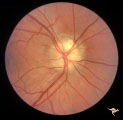 |
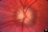
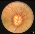
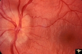
Visible Drusen | PP24a. Right eye. Exposed drusen. There are inferior nerve fiber layer defects in the upper arcuate bundles. Optic disc is also hypoplastic. Anatomy: Optic disc. Pathology: Drusen of the optic disc. Disease/Diagnosis: Drusen of the optic disc. Clinical: Hypoplastic optic disc with drusen. | Image |
| 27 |
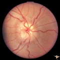 |
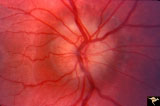
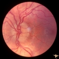
Visible Drusen - Bilateral | PP22a: right eye with obvious exposed drusen. PP22 b: Note bypass vein draining into the choroid at 8:00. Anatomy: Optic disc. Pathology: Drusen of the optic disc. Disease/Diagnosis: Drusen of the optic disc. Clinical: Normally functioning eye with drusen. | Image |
| 28 |
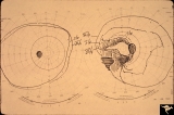 |
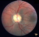
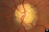
Visible Drusen with Visual Field Loss | 16 year old girl: Drusen disc. Goldmann visual field. Anatomy: Optic disc. Pathology: Drusen of the optic disc. Disease/Diagnosis: Drusen of the optic disc. Clinical: Drusen disc with visual field loss. | Image |
| 29 |
 |
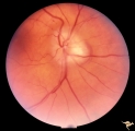
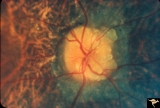
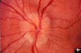
Visible Drusen with Visual Field Loss | PP25b right eye with drusen and severe visual field loss. Match with PP25a, c & d. Anatomy: Optic disc. Pathology: Drusen of the optic disc. Disease/Diagnosis: Drusen of the optic disc. Clinical: Drusen disc with servere visual field loss. | Image |
| 30 |
 |
Drusen Plus Papilledema | PP37a: right swollen disc on top of drusen with narrowing of the arterioles;PP37 b: left visible drusen and papilledema with sub-retinal hemorrhage temporally. Patient had frontal glioblastoma. Anatomy: Optic disc. Pathology: Drusen of the optic disc. Disease/Diagnosis: Drusen of the optic disc. Cli... | Image |
| 31 |
 |
Hemorrhagic Complication of Drusen | PP31a, left and PP31, right taken in April. PP31c: left taken after an interval of 2 months. Hemorrhage. Hemorrhagic complications of drusen. 15 year old boy. Anatomy: Optic disc. Pathology: Drusen of the optic disc. Disease/Diagnosis: Drusen of the optic disc. Clinical: Hemorrhage in drusen disc. | Image |
| 32 |
 |
Hemorrhagic Complication of Drusen | PP31a, left and PP31, right taken in April. PP31c: left taken after an interval of 2 months. Hemorrhage. Hemorrhagic complications of drusen. 15 year old boy. Anatomy: Optic disc. Pathology: Drusen of the optic disc. Disease/Diagnosis: Drusen of the optic disc. Clinical: Drusen. | Image |
| 33 |
 |
Visible Drusen with Visual Field Loss | PP25a: Left eye: Severe visual field defect. PP25b: right eye with exposed drusen and field loss: visual field defects; PP25c: right eye visual field PP25d: left eye visual field. Anatomy: Optic disc. Pathology: Drusen of the optic disc. Disease/Diagnosis: Drusen of the optic disc. Clinical: Dr... | Image |
| 34 |
 |
Buried Drusen | Young woman with pseudo papilledema from buried drusen with associated visual field defects. Barely visible in the upper arcuate nerve fibers is a slit like defect. Anatomy: Optic disc. Pathology: Drusen of the optic disc. Disease/Diagnosis: Drusen of the optic disc. Clinical notes: This patient had... | Image |
| 35 |
 |
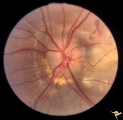
Visible Drusen with Retinitis Pigmentosa | Right eye. Optic disc drusen with retinitis pigmentosa. Note the marked narrowing of the retinal arterioles and the spectacular change in the peripapillary choroid. Anatomy: Optic disc. Pathology: Drusen of the optic disc. Disease/Diagnosis: Drusen of the optic disc. Clinical: Patient was nearly bli... | Image |
| 36 |
 |
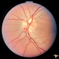
Buried Drusen | 7 year old boy with pseudo papilledema from buried drusen. Note the lumpy contour of the disc margin. Also note the surrounding ring-like light reflex that is optically perfect and indicates absence of edema spreading onto the surrounding retina. Anatomy: Optic disc. Pathology: Drusen of the optic d... | Image |
| 37 |
 |
Buried Drusen | Left disc has a blurred lumpy margin. Retinal vessels are not obscured in the disc margin blur, therefore no edema is present. This is an example of a difficult blurred disc, the nature of which is clarified by the presence of a clear cut disk anomoly in the fellow eye. 8 year old girl. PP_15a has b... | Image |
| 38 |
 |
Vascular Complications of Drusen: Drusen Causing Loss of Superior Retinal Arterial Supply | PP32a: right; PP32b: left eye. Left eye has occlusion of superior branch of the central retinal artery at 11:30 with the inferior retinal artery supplying collateral to the superior retina. Notice the branch of the inferior retinal artery moves superiorly heading toward the upper retina. Drusen w... | Image |
| 39 |
 |
Buried Drusen with Choroidal Retinal Scar | Right eye: Buried drusen; probable complication of peripapillary hemorrhage at 7:00. Anatomy: Optic disc. Pathology: Drusen of the optic disc. Disease/Diagnosis: Drusen of the optic disc. Clinical notes: Enlarged blind spot. | Image |
| 40 |
 |
Drusen with Vertical Retinal Folds | PP36a & b: Both left eye: Buried drusen. Note vertical retinal folds. Anatomy: Optic disc. Pathology: Drusen of the optic disc. Disease/Diagnosis: Drusen of the optic disc. | Image |
| 41 |
 |
Drusen with Vertical Retinal Folds | PP36a & b:Both left eye: Buried drusen. Note vertical retinal folds. Anatomy: Optic disc. Pathology: Drusen of the optic disc. Disease/Diagnosis: Drusen of the optic disc. | Image |
| 42 |
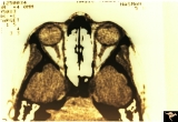 |
Unilateral Buried Drusen | PP23a: left eye; PP23b: CT scan showing calcium (bright spot at optic nerve head on left). 10 year old boy. Anatomy: Optic disc. Pathology: Drusen of the optic disc. Disease/Diagnosis: Drusen of the optic disc. Clinical: Unilateral drusen, only in the left eye. | Image |
| 43 |
 |
Visible Drusen - Bilateral | PP22a: right eye. PP22b: Note bypass vein draining into the choroid at 8:00. Anatomy: Optic disc. Pathology: Drusen of the optic disc. Disease/Diagnosis: Drusen of the optic disc. Clinical: Normally functioning eye with drusen. | Image |
| 44 |
 |
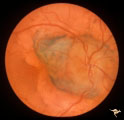
Buried Drusen with Sub-retinal Neovascular Net | Buried drusen with sub-retinal neovascular net. Both PP29a and PP29b are left eye: 17 year old girl; Visual acuity 10/400. Anatomy: Optic disc. Pathology: Drusen of the optic disc. Disease/Diagnosis: Drusen of the optic disc. Clinical notes: Loss of central vision due to subretinal neovascularizatio... | Image |
| 45 |
 |
Drusen with Sub-retinal Neovascular Net | Buried drusen with sub-retinal neovascular net. There may be retinoschisis as well. Anatomy: Optic disc. Pathology: Drusen plus neovascularization at the border of the optic disc. Disease/Diagnosis: Drusen of the optic disc. Clinical: Patient has very large blind spot and impaired central vision. | Image |
| 46 |
 |
Hemorrhagic Complication of Drusen | PP31a, left and PP31, right taken in April. PP31c: left taken after an interval of 2 months. Hemorrhage. Hemorrhagic complications of drusen. 15 year old boy. Anatomy: Optic disc. Pathology: Drusen of the optic disc. Disease/Diagnosis: Drusen of the optic disc. Clinical: Patient complained of blurre... | Image |
| 47 |
 |
Late Complications of Drusen | PP33a: right disc shows pallor and small calcified crystals on the disc surface. PP33: left disc shows calcified specs on temporal sector of the disc. Florid drusen in young patients changes over time to assume this appearance. Anatomy: Optic disc. Pathology: Drusen of the optic disc. Disease/Dia... | Image |
| 48 |
 |
Late Complications of Drusen | PP33a: right disc shows pallor and small calcified crystals on the disc surface. PP33b: left disc shows calcified specs on temporal sector of the disc. Florid drusen in young patients changes over time to assume this appearance. Anatomy: Optic disc. Pathology: Drusen of the optic disc. Disease/Di... | Image |
| 49 |
 |
Visible Drusen with Visual Field Loss | Right eye visual field combine with PP25a, b, & d. Anatomy: Optic disc. Pathology: Drusen of the optic disc. Disease/Diagnosis: Drusen of the optic disc. Clinical: Drusen disc with severe visual field defect. note the nasal visual field loss and the arcuate bundle defects. Central vision was 20/20. | Image |
| 50 |
 |
Buried Drusen with Sub-retinal Neovascular Net | Buried drusen with sub-retinal neovascular net. This is the same left eye. Appearance of the central retina of the left eye. Both PP29a & b are left eye: 17 year old girl; Visual acuity 10/400. Anatomy: Optic disc. Pathology: Drusen of the optic disc. DIsease/Diagnosis: Drusen of the optic disc. Cl... | Image |