Best known for his world-renowned neuro-ophthalmology unit based at the University of California, San Francisco, William Hoyt, MD collected here more than 850 of his best images covering a wide range of disorders.
William F. Hoyt, MD, Professor Emeritus of Ophthalmology, Neurology and Neurosurgery, Department of Ophthalmology, University of California, San Francisco.
NOVEL: https://novel.utah.edu/
TO
| Title | Description | Type | ||
|---|---|---|---|---|
| 426 |
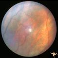 |
Von Hippel Lindau Disease | Von Hippel Lindau Disease. Retinal photograph showing small whitish hemangiomablastoma. Note the dilated arterial and venous channels entering and leaving the tumor. Anatomy: Retina. Pathology: Hemangioblastoma. Disease/Diagnosis: Von Hippel Lindau disease. Clinical: No visual symptoms. | Image |
| 427 |
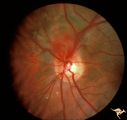 |
Von Hippel Lindau Disease (Hemangioblastoma of the Optic Disc) | Von Hippel Lindau Disease with a retinal hemangioblastoma on her optic disc. Anatomy: Optic disc. Pathology: Hemangioblastoma. Disease/Diagnosis: Von Hippel Lindau disease. Clinical: No visual symptoms. Patient had cerebellar ataxia. Imaging: R1_C1b is Arteriogram showing hemangioblastoma of the cer... | Image |
| 428 |
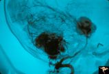 |
Von Hippel Lindau Disease (Hemangioblastoma of the Optic Disc) | Von Hippel Lindau Disease; Arteriogram showing hemangioblastoma of the cerebellum and midbrain. Anatomy: Brain. Pathology: Hemangioblastoma. Disease/Diagnosis: Von Hippel Lindau disease. Clinical: No visual symptoms. Patient had cerebellar ataxia. Imaging: Arteriogram showing hemangioblastoma of the... | Image |
| 429 |
 |
Von Hippel Lindau Disease (Retinal Hemangioblastoma) | Von Hippel Lindau Disease with large peripheral retinal hemangioblastoma. View of the tumor. Larger artery entering and the vein leaving the tumor are evidence of rapid arteriovenous shunting within the tumor. Group with R1_C3b, R1_C3a, R1_C3d. Anatomy: Retina. Pathology: Hemangioblastoma. Disease/... | Image |
| 430 |
 |
Von Hippel Lindau Disease (Retinal Hemangioblastoma) | Von Hippel Lindau Disease with large retinal hemangioblastoma. Continued view of the arteriole and venous channels leading to the tumor. Group with R1_C3a, R1_C3c, R1_C3d. Anatomy: Retina. Pathology: Hemangioblastoma. Disease/Diagnosis: Von Hippel Lindau disease. Clinical: No visual symptoms. | Image |
| 431 |
 |
Von Hippel Lindau Disease (Retinal Hemangioblastoma) | Von Hippel Lindau Disease (Retinal Hemangioblastoma); Small hemangioblastoma on the disc margin at 10:00. Large peripheral hemangioblastoma out of view to the top right seen on R1_C2b. Anatomy: Optic disc. Pathology: Hemangioblastoma. Disease/Diagnosis: Von Hippel Lindau disease. Clinical: No visual... | Image |
| 432 |
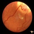 |
Von Hippel Lindau Disease (Retinal Hemangioblastoma) | Von Hippel Lindau Disease (Retinal Hemangioblastoma); Eye with large peripheral hemangioblastoma. Note particularly the large draining vein. Pair with R1_C2a. Anatomy: Optic disc. Pathology: Hemangioblastoma. Disease/Diagnosis: Von Hippel Lindau disease. Clinical: No visual symptoms. | Image |
| 433 |
 |
Von Hippel Lindau Disease (Retinal Hemangioblastoma) | Von Hippel Lindau Disease with large retinal hemangioblastoma. Huge arteriole and venous loops directed upward and nasally toward the tumor. Group with R1_C3b, R1_C3c, R1_C3d. Anatomy: Retina. Pathology: Hemangioblastoma. Disease/Diagnosis: Von Hippel Lindau disease. Clinical: No visual symptoms. | Image |
| 434 |
 |
Von Hippel Lindau Disease (Retinal Hemangioblastoma) | Von Hippel Lindau Disease with large peripheral retinal hemangioblastoma. View of the tumor. Larger artery entering and the vein leaving the tumor are evidence of rapid arteriovenous shunting within the tumor. Group with R1_C3b, R1_C3c, R1_C3c. Anatomy: Retina. Pathology: Hemangioblastoma. Disease/... | Image |
| 435 |
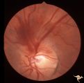 |
Von Hippel Lindau Disease Associated with Increased ICP | Von Hippel Lindau Disease; Optic disc lesion with hemorrhage from it in a patient with acute intracranial pressure elevation from a posterior fossa hemangioblastoma. Anatomy: Optic disc. Pathology: Hemangioblastoma. Disease/Diagnosis: Von Hippel Lindau disease. Clinical: Headache. | Image |
| 436 |
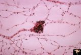 |
Von Hippel Lindau Disease Pathology of Mini Hemangioblastoma | Von Hippel Lindau Disease. Pathologic appearance of flat preparation of retina from necropsy study. Trypsin digestion study of retinal vascular bed with a mini VHL lesion. Anatomy: Retina. Pathology: Hemangioblastoma. Disease/Diagnosis: Von Hippel Lindau disease. Clinical: No visual symptoms. | Image |
| 437 |
 |
Von Hippel Lindau Disease with Photocoagulation Effect | Von Hippel Lindau Disease with appearance of Xenon photocoagulation on a mini hemangioblastoma. Anatomy: Retina. Pathology: Hemangioblastoma. Disease/Diagnosis: Von Hippel Lindau disease. Clinical: No visual symptoms. | Image |
