Best known for his world-renowned neuro-ophthalmology unit based at the University of California, San Francisco, William Hoyt, MD collected here more than 850 of his best images covering a wide range of disorders.
William F. Hoyt, MD, Professor Emeritus of Ophthalmology, Neurology and Neurosurgery, Department of Ophthalmology, University of California, San Francisco.
NOVEL: https://novel.utah.edu/
TO
| Title | Description | Type | ||
|---|---|---|---|---|
| 426 |
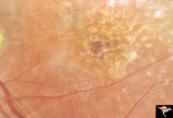 |
Pigmentary Retinopathy with Peripheral Neuropathy (Flecked Retinal Syndrome or Fundus Flavimaculatus) | Pigmentary retinopathy with peripheral neuropathy (Flecked Retinal Syndrome or Fundus Flavimaculatus) in a young woman. Anatomy: Retina. Pathology: Peripheral nerve degeneration. Disease/Diagnosis: Retinitis pigmentosa with hereditary peripheral degeneration. Clinical: Blindness. | Image |
| 427 |
 |
Pigmentary Retinopathy with Peripheral Neuropathy (Flecked Retinal Syndrome or Fundus Flavimaculatus) | Pigmentary retinopathy with peripheral neuropathy (Flecked Retinal Syndrome or Fundus Flavimaculatus) in a young woman. Anatomy: Retina. Pathology: Peripheral nerve degeneration. Disease/Diagnosis: Retinitis pigmentosa with hereditary peripheral degeneration. Clinical: Blindness. | Image |
| 428 |
 |
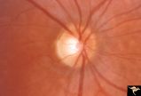
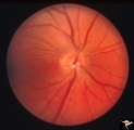
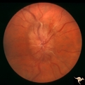
Post Papilledema | Right eye. Post Papilledema with minimal optic disc changes after treatment for temporal lobe glioma. Minimal optic disc haze. Optic disc. Pathology: Papilledema. Disease/Diagnosis: Post Papilledema due to temporal lobe glioma. | Image |
| 429 |
 |
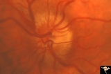
Post Papilledema | Left eye. Post Papilledema with minimal optic disc changes after treatment for temporal lobe glioma. Minimal optic disc haze. | Image |
| 430 |
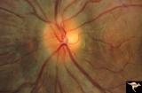 |
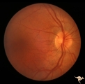
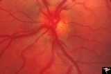
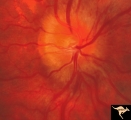
Post Papilledema Disc Blurring | Left eye. 8 year old boy. Post papilledema due to brain tumor. Note the entire peripapillary nerve fiber is blurred but the optic discs are barely elevated. Anatomy: Optic disc. Pathology: Brain tumor. Disease/Diagnosis: Papilledema. Clinical: Post papilledema due to brain tumor. | Image |
| 431 |
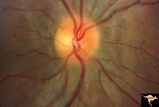 |
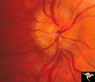
Post Papilledema Disc Blurring | Right eye. 8 year old boy. Post papilledema due to brain tumor. Note the entire peripapillary nerve fiber is blurred but the optic discs are barely elevated. Anatomy: Optic disc. Pathology: Post papilledema. Disease/Diagnosis: Post papilledema due to brain tumor. | Image |
| 432 |
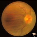 |
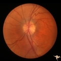
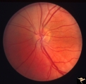
Post Papilledema with Choroidal Folds | Right eye. Post papilledema with choroidal folds due to brain tumor. Anatomy: Optic disc. Pathology: Post papilledema. Disease/Diagnosis: Post papilledema with choroidal folds. | Image |
| 433 |
 |
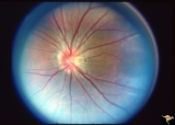
Post Papilledema, Secondary Optic Atrophy | Right eye. Post papilledema with chronic gliosis. arterial narrowing. ""high-water"" marks. Man. Anatomy: Optic disc. Pathology: Post papilledema. Disease/Diagnosis: Post papilledema with optic atrophy. | Image |
| 434 |
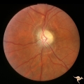 |
Post Papilledema, Secondary Optic Atrophy | Left eye. Post papilledema with chronic gliosis. arterial narrowing. "high-water" marks. Man. Anatomy: Optic disc. Pathology: Post papilledema. Disease/Diagnosis: Post papilledema with optic atrophy. | Image |
| 435 |
 |
PP5b Crowded Disc | PP5a: left eye;PP5 b: left eye X 2 magnification; congenital disc blurring. Boy. Anatomy: Optic disc. Pathology: Normal variation of the optic disc. Disease/ Diagnosis: Normal variation of the optic disc. Congenital blurred disc. Clinical: Blurred disc margin. Beautiful example of difficult differen... | Image |
| 436 |
 |
PP6a Crowded Disc with Glial Remnant | PP6a: 35 year old man. Right eye that has the glial remnant and blurred margins. PP6b: left eye. Anatomy: Optic disc. Pathology: Normal variation of the optic disc with glial remnant. Disease/ Diagnosis: Normal variation of the optic disc. Crowded disc with glial remnant. Clinical: Man referred for ... | Image |
| 437 |
 |
PP6b Crowded Disc with Glial Remnant | PP6a: right eye that has the glial remnant and blurred margins. PP6b: left eye. Anatomy: Optic disc. Pathology: Normal variation of the optic disc. Disease/ Diagnosis: Normal variation of the optic disc. Crowded disc. Clinical: Left eye is normal. | Image |
| 438 |
 |
PP7a Crowded disc | PP7a: right eye crowded disc with blurred margin. Note anomalous vascular pattern and glial tissue on the disc; PP7b- left disc is cupless disc and normal. 10 year old girl with gonadal dysgenesis and growth retardation. Anatomy: Optic disc. Pathology: Normal variation of the optic disc. Disease/ Di... | Image |
| 439 |
 |
PP8a Crowded Disc with Significant Nasal Disc Blurring | Congenital nasal disc blurring. Myopic eyes. Thai girl patient. One wonders about vitreal adherence to the disc. PP 8a right eye. Pair with left eye in PP8b. Anatomy: Optic disc. Pathology: Normal variation of the optic disc. Disease/ Diagnosis: Normal variation of the optic disc. Congenital blurre... | Image |
| 440 |
 |
PP8b Crowded Disc with Significant Nasal Disc Blurring | Congenital nasal disc blurring. Myopic eyes. Thai girl patient. One wonders about vitreal adherence to the disc. PP 8b left eye. Pair with PP 8a right eye. Anatomy: Optic disc. Pathology: Normal variation of the optic disc. Disease/ Diagnosis: Normal variation of the optic disc. Congenital blurred d... | Image |
| 441 |
 |
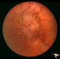
Progression of Papilledema due to Metastatic Melanoma | Right eye. Rapid progression of papilledema due to metastatic occipital melanoma. Papilladema has increased so that it has almost filled in the optic cup. Anatomy: Optic disc. Pathology: Papilledema. Disease/Diagnosis: Papilledema due to metastatic occipital melanoma. | Image |
| 442 |
 |
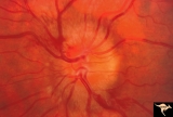
Progression of Papilledema due to Metastatic Melanoma | Left eye at presentation. Early stage. Rapid progression of papilledema due to metastatic occipital melanoma. Anatomy: Optic disc. Pathology: Papilledema. Disease/Diagnosis: Papilledema due to metastatic occipital melanoma. | Image |
| 443 |
 |
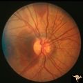
Progression of Papilledema due to Metastatic Melanoma | Right eye at presentation. Early stage bilateral papilledema in a man. Note increased papilledema. Rapid progression of papilledema due to occipital metastatic melanoma. Anatomy: Optic disc. Pathology: Papilledema. Disease/Diagnosis: Early stage bilateral papilledema. | Image |
| 444 |
 |
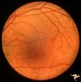
Progression of Papilledema due to Metastatic Melanoma | Right eye at presentation. Early stage bilateral papilledema in a man. Rapid progression of bilateral papilledema due to metastatic occipital melanoma. Anatomy: Optic disc. Pathology: Papilledema. Disease/Diagnosis: Papilledema. | Image |
| 445 |
 |
Progression of Papilledema due to Metastatic Melanoma | Left eye one and a half months after presentation. Papilledema has increased and now hemorrhages have been added. Rapid progression of papilledema due to metastatic occipital melanoma. | Image |
| 446 |
 |
Progression of Papilledema due to Metastatic Melanoma | Left eye. 10 days after presentation. Papilledema has increased and bleeding is occurring at the disc margins. Rapid progression of papilledema due to metastatic occipital melanoma. | Image |
| 447 |
 |
Pseudoxanthoma Elasticum (PXE) | Pseudoxanthoma elasticum (PXE) Right eye shows angiod streaks with associated hemorrhage. Patient was 25 year old man who developed a right sided carotid cavernous fistula. Anatomy: Retina. Pathology: Pseudoxanthoma elasticum (PXE). Disease/Diagnosis: PXE with angiod streaks with associated hemorrha... | Image |
| 448 |
 |
Pseudoxanthoma Elasticum (PXE) | Pseudoxanthoma elasticum (PXE). Left eye. Peripheral retina shows characteristic sign of PXE called Peau d'orange. Anatomy: Retina. Pathology: Pseudoxanthoma elasticum (PXE). Disease/Diagnosis: PXE with angiod streaks with associated hemorrhage associated with carotid cavernous fistula. Clinical: Vi... | Image |
| 449 |
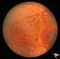 |
Pseudoxanthoma Elasticum (PXE) | Pseudoxanthoma elasticum (PXE). Right eye. Peripheral retina shows characteristic sign of PXE called Peau d'orange. Anatomy: Retina. Pathology: Pseudoxanthoma elasticum (PXE). Disease/Diagnosis: PXE with angiod streaks with associated hemorrhage associated with carotid cavernous fistula. Clinical: V... | Image |
| 450 |
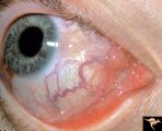 |
Pseudoxanthoma Elasticum (PXE) | Pseudoxanthoma elasticum (PXE) Conjunctival signs of carotid cavernous fistula of the right eye. Anatomy: Retina. Pathology: Pseudoxanthoma elasticum (PXE). Disease/Diagnosis: PXE with angiod streaks with associated hemorrhage associated with carotid cavernous fistula. Clinical: Vision blurred in ri... | Image |