Best known for his world-renowned neuro-ophthalmology unit based at the University of California, San Francisco, William Hoyt, MD collected here more than 850 of his best images covering a wide range of disorders.
William F. Hoyt, MD, Professor Emeritus of Ophthalmology, Neurology and Neurosurgery, Department of Ophthalmology, University of California, San Francisco.
NOVEL: https://novel.utah.edu/
TO
| Title | Description | Type | ||
|---|---|---|---|---|
| 376 |
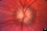 |
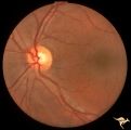
Hemorrhagic Complication of Drusen | PP31a, left and PP31, right taken in April. PP31c: left taken after an interval of 2 months. Hemorrhage. Hemorrhagic complications of drusen. 15 year old boy. Anatomy: Optic disc. Pathology: Drusen of the optic disc. Disease/Diagnosis: Drusen of the optic disc. Clinical: Hemorrhage in drusen disc. | Image |
| 377 |
 |
Hemorrhagic Complication of Drusen | PP31a, left and PP31, right taken in April. PP31c: left taken after an interval of 2 months. Hemorrhage. Hemorrhagic complications of drusen. 15 year old boy. Anatomy: Optic disc. Pathology: Drusen of the optic disc. Disease/Diagnosis: Drusen of the optic disc. Clinical: Drusen. | Image |
| 378 |
 |
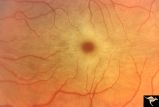
Hereditary Macular Degenerative Disease with Spastic Paraplegia | Hereditary macular degenerative disease with Patient has spastic paraplegia associated with hereditary macular degenerative disease. Anatomy: Retina. Pathology: Cerebellar spinal degenerative disease. Disease/Diagnosis: Retinitis pigmentosa with spinal degeneration. Clinical: Hereditary spastic para... | Image |
| 379 |
 |
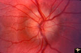
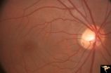
Ischemic Complication of Drusen | PP30a: right eye--buried drusen; PP30b: buried drusen with anterior ischemic optic neuropathy (AION) from complication of drusen of left eye. Ischemic complication of drusen in left eye. PP30c: 3 month follow-up: narrowed arterioles slightly pale disc with buried drusen. Anatomy: Optic disc. Patho... | Image |
| 380 |
 |
Ischemic Complication of Drusen | PP30a: right eye--buried drusen; PP3-b: buried drusen with anterior ischemic optic neuropathy (AION) from complication of drusen of left eye. Ischemic complication of drusen in left eye. PP30c: 3 month follow-up: narrowed arterioles slightly pale disc with buried drusen. Anatomy: Optic disc. Path... | Image |
| 381 |
 |
Ischemic Complication of Drusen | PP30a: right eye--buried drusen; PP30b: buried drusen with anterior ischemic optic neuropathy (AION) from complication of drusen of left eye. Ischemic complication of drusen in left eye. PP30c: 3 month follow-up: narrowed arterioles slightly pale disc with buried drusen. Anatomy: Optic disc. Patho... | Image |
| 382 |
 |
Late Complications of Drusen | PP33a: right disc shows pallor and small calcified crystals on the disc surface. PP33: left disc shows calcified specs on temporal sector of the disc. Florid drusen in young patients changes over time to assume this appearance. Anatomy: Optic disc. Pathology: Drusen of the optic disc. Disease/Dia... | Image |
| 383 |
 |
Late Complications of Drusen | PP33a: right disc shows pallor and small calcified crystals on the disc surface. PP33b: left disc shows calcified specs on temporal sector of the disc. Florid drusen in young patients changes over time to assume this appearance. Anatomy: Optic disc. Pathology: Drusen of the optic disc. Disease/Di... | Image |
| 384 |
 |
Macular Cherry Red Spots in Niemann-Pick disease | Close up view of macular cherry red spots in Niemann-Pick disease. Same patient as R2A2a. Anatomy: Retina. Pathology: Retinal ganglion cell accumulation of lipid. Disease/Diagnosis: Niemann-Pick disease. Clinical: Severe mental retardation and blindness. Fatal. | Image |
| 385 |
 |
Macular Cherry Red Spots in Niemann-Pick disease | Macular cherry red spots in Niemann-Pick disease. Same patient as R2A2b. Anatomy: Retina. Pathology: Retinal ganglion cell accumulation of lipid. Disease/Diagnosis: Niemann-Pick disease. Clinical: Severe mental retardation and blindness. Fatal. | Image |
| 386 |
 |
Macular Cherry Red Spots in Tay-Sachs disease | Macular cherry red spots in patient with Tay-Sachs disease. Anatomy: Retina. Pathology: Retinal ganglion cell accumulation of lipid. Disease/Diagnosis: Tay-Sachs disease. Clinical: Severe mental retardation and blindness. Fatal. | Image |
| 387 |
 |
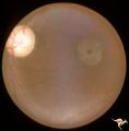
Medullated Nerve Fibers with Papilledema | Left eye. papilledema only. Man with metastatic gastric carcinoma. Anatomy: Optic disc. Pathology: Papilledema. Disease/Diagnosis: Papilledema plus medullated nerve fibers. | Image |
| 388 |
 |
Medullated Nerve Fibers with Papilledema | Right eye. Papilledema superimposed upon medullated nerve fibers. Man with metastatic gastric carcinoma. Anatomy: Optic disc. Pathology: Papilledema. Disease/Diagnosis: Papilledema plus medullated nerve fibers. | Image |
| 389 |
 |
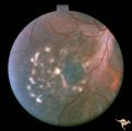
Multifocal Choroidopathy | Multifocal choroidopathy in a patient with uveitis. Anatomy: Retina. Disease/Diagnosis: Uveitis, Multifocal placoid pigment epitheliopathy. Clinical: Visual loss. | Image |
| 390 |
 |
Multifocal Choroidopathy | Multifocal choroidopathy in a patient with uveitis. Anatomy: Retina. Disease/Diagnosis: Uveitis, Multifocal placoid pigment epitheliopathy. Clinical: Visual loss. | Image |
| 391 |
 |
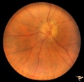
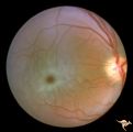
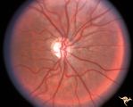
Neurofibromatosis-1 | Extensive retinal microvascular malformation involving both small and large retinal vessels. (Ref: BJO 2002:86, p282-284). Anatomy: Retina. Pathology: Retinal microvascular malformations. Disease/Diagnosis: Neurofibromatosis type 1. Clinical: No visual symptoms. | Image |
| 392 |
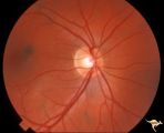 |
Neurofibromatosis-1 | Normal appearing optic disc with dark pigmented choroidal nevi. The patient had NF-1 and had a subclinical optic glioma on the left eye. This is the right eye. Anatomy: Optic disc. Pathology: Choroidal nevus. Disease/Diagnosis: Neurofibromatosis type 1. Clinical: No visual symptoms. | Image |
| 393 |
 |
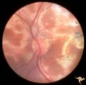
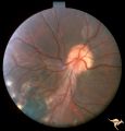
Neurofibromatosis-1 | Optic atrophy and hypoplasia of the optic disc associated with chiasmal glioma in a patient with NF-1. Anatomy: Optic disc. Pathology: Chiasmal glioma; Optic atrophy; Hypoplasia. Disease/Diagnosis: Neurofibromatosis type 1. Clinical: Proptosis; Blindness. | Image |
| 394 |
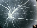 |
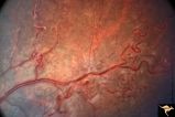
Neurofibromatosis-1 | Fluorescein angiogram defines the extent of the microvascular malformation. Pair with R1_E5b. (Ref: BJO 2002:86, p282-284). Anatomy: Retina. Pathology: Retinal microvascular malformations. Disease/Diagnosis: Neurofibromatosis type 1. Clinical: No visual symptoms. Imaging: Fluorescein angiogram. | Image |
| 395 |
 |
Neurofibromatosis-1 | Retinal microvascular malformations in NF-1. Fundus picture shows a somewhat larger vertically running corkscrew malformation between two temporal retinal veins. Pair with R1_E5a. Anatomy: Retina. Pathology: Retinal microvascular malformations. Disease/Diagnosis: Neurofibromatosis type 1. Clinical: ... | Image |
| 396 |
 |
Neurofibromatosis-1 | Retinal microvascular malformations in NF-1 located between the disc and the macula. Anatomy: Retina. Pathology: Retinal microvascular malformations. Disease/Diagnosis: Neurofibromatosis type 1. Clinical: No visual symptoms. | Image |
| 397 |
 |
Neurofibromatosis-1 | Retinal microvascular malformations between optic disc and macula in NF-1. Anatomy: Retina. Pathology: Retinal microvascular malformations. Disease/Diagnosis: Neurofibromatosis type 1. Clinical: No visual symptoms. | Image |
| 398 |
 |
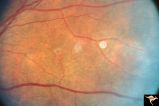
Neurofibromatosis-2 | CPERH (choroidal pigment epithelial retinal hamartoma) lesion in a patient with NF-2. Note the oblique superficial retinal traction folds running toward the center of the main lesion. 51 year old man. Anatomy: Retina. Pathology: Hamartoma. Disease/Diagnosis: Neurofibromatosis type 2. Clinical: Fiel... | Image |
| 399 |
 |
Neurofibromatosis-2 | Retinal tumor in NF-2 referred to as a CPERH (choroidal pigment epithelial retinal hamartoma). Patient, a 16 year old girl, had bilateral acoustic neurinomas. Pair with R1_F2b. Same eye. Anatomy: Optic disc; Retina. Pathology: Retinal hamartoma; Bilateral acoustic neurinoma. Disease/Diagnosis: Neuro... | Image |
| 400 |
 |
Neurofibromatosis-2 | Retinal tumor in NF-2 referred to as a CPERH (choroidal pigment epithelial retinal hamartoma). Patient, a 16 year old girl, had bilateral acoustic neurinomas. Pair with R1_F2a. Same eye. Anatomy: Optic disc; Retina. Pathology: Retinal hamartoma; Bilateral acoustic neurinoma. Disease/Diagnosis: Neuro... | Image |