Best known for his world-renowned neuro-ophthalmology unit based at the University of California, San Francisco, William Hoyt, MD collected here more than 850 of his best images covering a wide range of disorders.
William F. Hoyt, MD, Professor Emeritus of Ophthalmology, Neurology and Neurosurgery, Department of Ophthalmology, University of California, San Francisco.
NOVEL: https://novel.utah.edu/
TO
| Title | Description | Type | ||
|---|---|---|---|---|
| 351 |
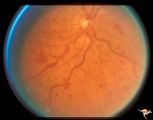 |
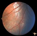
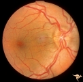
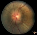
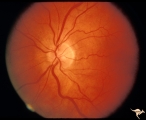
Slow Flow (Chronic Hypoxic) Retinopathy | Examples of Slow flow (chronic hypoxic) retinopathy showing dilated and tortuous retinal veins and multiple capillary hemorrhages. This kind of retinopathy is produced by impaired arteriole circulation to the retina from various causes. Anatomy: Retina. Pathology: Ophthalmic artery venous malformati... | Image |
| 352 |
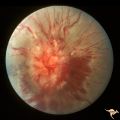 |
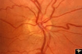
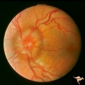
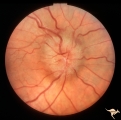
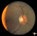
Sturge Weber Syndrome (Encephalotrigeminal Angiomatosis) | Sturge Weber Syndrome (Encephalotrigeminal angiomatosis) with retinal evidence of central retinal vein occlusion. Anatomy: Retina. Pathology: Diffuse choroidal hemangioma; Glaucoma. Disease/Diagnosis: Sturge Weber Syndrome. Clinical: Port wine hemangioma of the face. | Image |
| 353 |
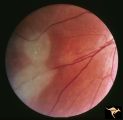 |
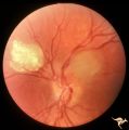
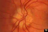
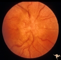
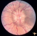
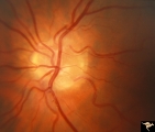
Tuberous Sclerosis | Tuberous Sclerosis. Has two astrocytic hamartomas. Lower shows a few specs of calcification. Note how the tumor obscures the underlying retinal vessels. Left eye. Pair with R1_D4a. Anatomy: Retina. Pathology: Astrocytic hamartoma. Disease/Diagnosis: Tuberous sclerosis. Clinical: No visual symptoms. | Image |
| 354 |
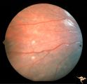 |
Tuberous Sclerosis | Two very small tuberous sclerosis lesions. Group with R1_D7a, b, d. Anatomy: Retina. Pathology: Astrocytic hamartoma. Disease/Diagnosis: Tuberous sclerosis. Clinical: Patient had mental retardation and epilepsy. No visual symptoms. | Image |
| 355 |
 |
Tuberous Sclerosis | Tuberous Sclerosis. Has two astrocytic hamartomas. One is calcified. Right eye. Pair with R1_D4b. Anatomy: Optic disc; Retina. Pathology: Astrocytic hamartoma. Disease/Diagnosis: Tuberous sclerosis. Clinical: No visual symptoms. | Image |
| 356 |
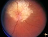 |
Tuberous Sclerosis | Tuberous Sclerosis. Astrocytic hamartoma of the optic disc. Anatomy: Optic disc. Pathology: Astrocytic hamartoma. Disease/Diagnosis: Tuberous sclerosis. Clinical: No visual symptoms. | Image |
| 357 |
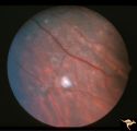 |
Tuberous Sclerosis | Several small tuberous sclerosis lesions. Group with R1_D7a, c, d. Anatomy: Retina. Pathology: Astrocytic hamartoma. Disease/Diagnosis: Tuberous sclerosis. Clinical: Patient had mental retardation and epilepsy. No visual symptoms. | Image |
| 358 |
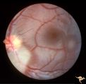 |
Tuberous Sclerosis | Two moderate sized tuberous sclerosis lesions in the left eye. Group with R1_D7b, c, d. Anatomy: Optic disc; Retina. Pathology: Astrocytic hamartoma. Disease/Diagnosis: Tuberous sclerosis. Clinical: Patient had mental retardation and epilepsy. No visual symptoms. | Image |
| 359 |
 |
Tuberous Sclerosis | One small tuberous sclerosis lesion. Group with R1_D7a, b, c. Anatomy: Retina. Pathology: Astrocytic hamartoma. Disease/Diagnosis: Tuberous sclerosis. Clinical: Patient had mental retardation and epilepsy. No visual symptoms. | Image |
| 360 |
 |
Unilateral Buried Drusen | PP20a: Right eye. Normal disc without optic cup.PP20b: Left eye. Buried drusen nasally and exposed drusen at the temporal margin. Boy. Anatomy: Optic disc. Pathology: Drusen of the optic disc. Disease/Diagnosis: Drusen of the optic disc. Clinical: Normally functioning eye with drusen. Right eye nor... | Image |
| 361 |
 |
Unilateral Buried Drusen | PP20a: Right eye. Normal disc without optic cup.PP20b: Left eye. Buried drusen nasally and exposed drusen at the temporal margin. Boy. Anatomy: Optic disc. Pathology: Drusen of the optic disc.. Disease/Diagnosis: Drusen of the optic disc.. Clinical: Normally functioning eye with drusen. Right eye n... | Image |
| 362 |
 |
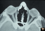
Unilateral Buried Drusen | PP23a: left eye; lumpy disc with buried drusen. PP23b: CT scan showing calcium (bright spot at optic nerve head on left). 10 year old boy. Anatomy: Optic disc. Pathology: Drusen of the optic disc. Disease/Diagnosis: Drusen of the optic disc. Clinical: Normally functioning eye with drusen. | Image |
| 363 |
 |
Unilateral Buried Drusen | PP23a: left eye; PP23b: CT scan showing calcium (bright spot at optic nerve head on left). 10 year old boy. Anatomy: Optic disc. Pathology: Drusen of the optic disc. Disease/Diagnosis: Drusen of the optic disc. Clinical: Unilateral drusen, only in the left eye. | Image |
| 364 |
 |
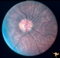
Unilateral Papilledema | Unilateral papilledema in Pseudotumor cerebri. Right eye. Has no cup. Woman. Anatomy: Optic disc. Pathology: Unilateral papilledema. Disease/Diagnosis: Idiopathic intracranial hypertension, pseudotumor cerebri. Clinical: Woman, headache. | Image |
| 365 |
 |
Unilateral Papilledema | Left eye. Has chronic papilledema. Woman. Anatomy: Optic disc. Pathology: Unilateral papilledema. Disease/Diagnosis: Idiopathic intracranial hypertension, pseudotumor cerebri. Clinical: Woman, headache. | Image |
| 366 |
 |
Unilateral Papilledema | Right eye.Chronic Papilledema in right eye. Woman. Pseudo Foster Kennedy due to asymmetric papilledema. Anatomy: Optic disc. Pathology: Chronic papilledema; optic atrophy. Disease/Diagnosis: Idiopathic intracranial hypertension (pseudotumor cerebri) causing pseudo Foster-Kennedy Syndrome. Clinical: ... | Image |
| 367 |
 |
Unilateral Papilledema | Left eye. Has no optic cup. 24 year old obese woman. Anatomy: Optic disc. Pathology: Unilateral papilledema. Disease/Diagnosis: Idiopthatic intracranial hypertension, pseudotumor cerebri. Clinical: Woman, headache, transient visual obscurations. | Image |
| 368 |
 |
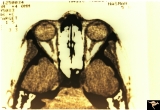
Unilateral Papilledema | CT scan showing equal thickening of the optic nerves. CT scan of no help in determining which eye has the papilledema. In this case, right eye has papilledema. Scan of patient depicted P_13a and P_13b. Anatomy: Optic disc. Pathology: Unilateral papilledema. Disease/Diagnosis: Idiopathic intracranial... | Image |
| 369 |
 |
Unilateral Papilledema | Left eye. Patient had tumor on right side. Right sided large meningioma. optociliary shunt at 10:00. Foster Kennedy. Anatomy: Optic disc. Pathology: Unilateral papilledema. Disease/Diagnosis: Meningioma of the brain. Clinical: Headache. | Image |
| 370 |
 |
Unilateral Papilledema | Right eye. Has papilledema. Patient has pseudotumor cerebri. 24 year old obese woman. Anatomy: Optic disc. Pathology: Unilateral papilledema. Disease/Diagnosis: Idiopathic intracranial hypertension, pseudotumor cerebri. Clinical: Woman, headache, transient visual obscurations. | Image |
| 371 |
 |
Unilateral Papilledema | Left eye. Has papilledema. 27 year old white woman. Anatomy: Optic disc. Pathology: Unilateral papilledema. Disease/Diagnosis: Idiopathic intracranial hypertension, pseudotumor cerebri. Clinical: Woman, headache. | Image |
| 372 |
 |
Unilateral Papilledema | Unilateral papilledema in pseudotumor cerebri. Right eye. Has no cup. 27 year old white woman. Anatomy: Optic disc. Pathology: Unilateral papilledema. Disease/Diagnosis: Idiopathic intracranial hypertiension, pseudotumor cerebri. Clinical: Woman, headache. | Image |
| 373 |
 |
Unilateral Papilledema | Left eye. Flat cupless disc. Woman. Anatomy: Optic disc. Pathology: Chronic papilledema; optic atrophy. Disease/Diagnosis: Idiopathic intracranial hypertension (pseudotumor cerebri) causing pseudo Foster-Kennedy Syndrome. Clinical: Visual loss in atrophic eye; obese. | Image |
| 374 |
 |
Unilateral Papilledema | Right eye. Patient had tumor on right side. Right sided large meningioma. Disc edema due to tumor. 29 year old black woman. The right disc has mild temporal pallor. Anatomy: Optic disc. Pathology: Uninaleral papilledema. Disease/Diagnosis: Meningioma of the brain. Clinical: Headache. | Image |
| 375 |
 |
Vascular Complications of Drusen | PP34a: Right eye. Superior retinal vein drains into the choroid at 12:00. It has occluded between center of disc and 12:00. Note white ghost vessel. Note that other veins drain into the disc edge at 4:00. There is no evidence of a central retinal vein in the middle of the disc. PP34b: Visible drus... | Image |