Best known for his world-renowned neuro-ophthalmology unit based at the University of California, San Francisco, William Hoyt, MD collected here more than 850 of his best images covering a wide range of disorders.
William F. Hoyt, MD, Professor Emeritus of Ophthalmology, Neurology and Neurosurgery, Department of Ophthalmology, University of California, San Francisco.
NOVEL: https://novel.utah.edu/
TO
| Title | Description | Type | ||
|---|---|---|---|---|
| 351 |
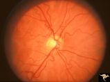 |
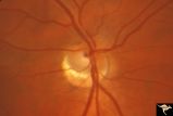
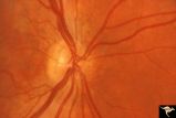
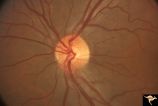
H73 Superior Segmental Optic Hypoplasia (SSOH) Topless Disc Syndrome | Bilateral SSOH. Left eye. Same patient as H_73. Anatomy: Optic disc. Pathology: Superior segmental optic hypoplasia (SSOH). Disease/ Diagnosis: Congenital anomaly. | Image |
| 352 |
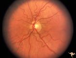 |
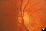
H74 Superior Segmental Optic Hypoplasia (SSOH) Topless Disc Syndrome | Bilateral SSOH. Right eye. Same patient as H_74. Anatomy: Optic disc. Pathology: Superior segmental optic hypoplasia (SSOH). Disease/ Diagnosis: Congenital anomaly. | Image |
| 353 |
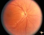 |
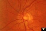
H75 Superior Segmental Optic Hypoplasia (SSOH) Topless Disc Syndrome | SSOH with hypoplasia of whole disc. Right eye. Anatomy: Optic disc. Pathology: Superior segmental optic hypoplasia (SSOH). Disease/ Diagnosis: Congenital anomaly. | Image |
| 354 |
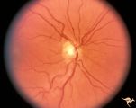 |
H76 Superior Segmental Optic Hypoplasia (SSOH) Topless Disc Syndrome | SSOH. Right eye. Anatomy: Optic disc. Pathology: Superior segmental optic hypoplasia (SSOH). Disease/ Diagnosis: Congenital anomaly. | Image |
| 355 |
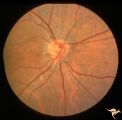 |
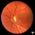
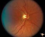
H77 Inferior Segmental Optic Hypoplasia (ISOH) | ISOH. Superior visual field defect. Inferior choroidal crescent. Anatomy: Optic disc. Pathology: Inferior segmental optic hypoplasia (ISOH). Disease/ Diagnosis: Congenital anomaly. | Image |
| 356 |
 |
H78 Inferior Segmental Optic Hypoplasia (ISOH) | ISOH with inferior choroidal crescent. Patient had superior visual field defect. Anatomy: Optic disc. Pathology: Inferior segmental optic hypoplasia (ISOH). Disease/ Diagnosis: Congenital anomaly. | Image |
| 357 |
 |
H79 Inferior Segmental Optic Hypoplasia (ISOH) | ISOH. Anatomy: Optic disc. Pathology: Inferior segmental optic hypoplasia (ISOH). Disease/ Diagnosis: Congenital anomaly. | Image |
| 358 |
 |
H80 Chiasmal Hemioptic Hypoplasia | Discs show striking nasal hypoplasia and band atrophy. DeMorsier synrome. Congenital bitemporal hemianopia with see-saw nystagmus. Note vertically oral shape of these hypoplastic nerves. The CT scan showed the median bar of the chiasm in this patient is totally hypoplastic. Anatomy: Optic disc. Path... | Image |
| 359 |
 |
H81 Chiasmal Hemioptic Hypoplasia | De Morsier synrome with congenital bitemporal hemianopia. Right eye. Note nasal hypoplasia of the right optic disc. Same patient as H_82. Anatomy: Optic disc. Pathology: Chiasmal hemioptic hypoplasia. Disease/ Diagnosis: Congenital anomaly involving chiasm | Image |
| 360 |
 |
H82 Chiasmal Hemioptic Hypoplasia | De Morsier synrome with congenital bitemporal hemianopia. Left eye. Same patient as H_81. Anatomy: Optic disc. Pathology: Chiasmal hemioptic hypoplasia. Disease/ Diagnosis: Congenital anomaly involving chiasm. | Image |
| 361 |
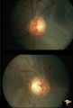 |
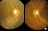
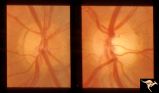
H83 Chiasmal Hemioptic Hypoplasia | De Morsier synrome with congenital bitemporal hemianopia. Note nasal hypoplasia of both optic discs. Left eye above, right eye below. Anatomy: Optic disc. Pathology: Chiasmal hemioptic hypoplasia. Disease/ Diagnosis: Congenital anomaly involving chiasm. | Image |
| 362 |
 |
H84 Chiasmal Hemioptic Hypoplasia | Congenital bitemporal hemianopia with marked bi-nasal hypoplasia. Left eye. 17 year old male. Same patient as H_85. Anatomy: Optic disc. Pathology: Chiasmal hemioptic hypoplasia. Disease/ Diagnosis: Congenital anomaly involving chiasm. | Image |
| 363 |
 |
H85 Chiasmal Hemioptic Hypoplasia | Congenital bitemporal hemianopia with marked bi-nasal hypoplasia. Right eye. 17 year old male. Same patient as H_84. Anatomy: Optic disc. Pathology: Chiasmal hemioptic hypoplasia. Disease/ Diagnosis: Congenital anomaly involving chiasm. | Image |
| 364 |
 |
H86 Chiasmal Hemioptic Hypoplasia | Congenital bitemporal hemianopia with nasal hypoplasia. 24 year old man. Same patient as H_87. Anatomy: Optic disc. Pathology: Chiasmal hemioptic hypoplasia. Disease/ Diagnosis: Congenital anomaly involving chiasm. | Image |
| 365 |
 |
H87 Chiasmal Hemioptic Hypoplasia | Congenital bitemporal hemianopia with nasal hypoplasia. 24 year old man. Same patient as H_86. Anatomy: Optic disc. Pathology: Chiasmal hemioptic hypoplasia. Disease/ Diagnosis: Congenital anomaly involving chiasm. | Image |
| 366 |
 |
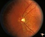
H88 Chiasmal Hemioptic Hypoplasia | Nasal hypoplasia with temporal hemianopia from a congenital Rathke Pouch Cyst. Anatomy: Optic disc. Pathology: Chiasmal hemioptic hypoplasia. Disease/ Diagnosis: Congenital anomaly involving chiasm. | Image |
| 367 |
 |
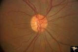
H90 Occipital Hemianoptic Hypoplasia | Note left disc (right side of image) is the eye with temporal field defect. Shows band atrophy. Anatomy: Optic disc. Pathology: Occipital hemianoptic hypoplasia. Congenital defect of the occipital lobe. | Image |
| 368 |
 |
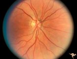
H91 Occipital Hemianoptic Hypoplasia | Left eye with temporal field defect shows trans-synaptic band atrophy. Same patient as H_92. Anatomy: Optic disc. Pathology: Occipital hemianoptic hypoplasia. Disease/ Diagnosis: Congenital defect of the occipital lobe. | Image |
| 369 |
 |
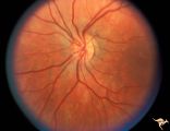
H92 Occipital Hemianoptic Hypoplasia | Right eye. Same patient as H_91. Anatomy: Optic disc. Pathology: Occipital hemianoptic hypoplasia. Disease/ Diagnosis: Congenital defect of the occipital lobe. | Image |
| 370 |
 |
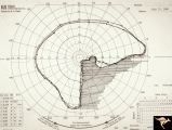
H93 Occipital Hemianoptic Hypoplasia | Visual field. Left eye. Right inferior homonymous. Same patient as H_94, H_95, H_96, H_97. Anatomy: Optic disc. Pathology: Occipital hemianoptic hypoplasia. Disease/ Diagnosis: Congenital defect of the occipital lobe. Imaging: MRI scan - See slide H97. | Image |
| 371 |
 |
H94 Occipital Hemianoptic Hypoplasia | Visual field. Right eye. Quatrantanopia. Same patient as H_93, H_95, H_96, H_97. Anatomy: Optic disc. Pathology: Occipital hemianoptic hypoplasia. Disease/ Diagnosis: Congenital defect of the occipital lobe. Imaging: MRI scan - See slide H97. | Image |
| 372 |
 |
H95 Occipital Hemianoptic Hypoplasia | Right eye with temporal field defect shows trans-synaptic band atrophy. Absence of nasal nerve fibers. Same patient as H_93, H_94, H_96, H_97. Anatomy: Optic disc. Pathology: Occipital hemianoptic hypoplasia. Disease/ Diagnosis: Congenital defect of the occipital lobe. Imaging: MRI scan - See slide ... | Image |
| 373 |
 |
H96 Occipital Hemianoptic Hypoplasia | Left eye has nasal quadrantic field defect. Same patient as H_93, H_94, H_95, H_97. Anatomy: Optic disc. Pathology: Occipital hemianoptic hypoplasia. Disease/ Diagnosis: Congenital defect of the occipital lobe. Imaging: MRI scan - See slide H97. | Image |
| 374 |
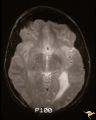 |
H97 Occipital Hemianoptic Hypoplasia | MRI scan shows left occipital lobe periventricular leuko-melacia. Same patient as H_93, H_94, H_95, H_96. Anatomy: Optic disc. Pathology: Occipital hemianoptic hypoplasia. DIsease/ Diagnosis: Congenital defect of the occipital lobe. Imaging: MRI scan. | Image |
| 375 |
 |
Hemorrhagic Complication of Drusen | PP31a, left and PP31, right taken in April. PP31c: left taken after an interval of 2 months. Hemorrhage. Hemorrhagic complications of drusen. 15 year old boy. Anatomy: Optic disc. Pathology: Drusen of the optic disc. Disease/Diagnosis: Drusen of the optic disc. Clinical: Patient complained of blurre... | Image |