Best known for his world-renowned neuro-ophthalmology unit based at the University of California, San Francisco, William Hoyt, MD collected here more than 850 of his best images covering a wide range of disorders.
William F. Hoyt, MD, Professor Emeritus of Ophthalmology, Neurology and Neurosurgery, Department of Ophthalmology, University of California, San Francisco.
NOVEL: https://novel.utah.edu/
TO
| Title | Description | Type | ||
|---|---|---|---|---|
| 301 |
 |
Neurofibromatosis-2 | CPERH (choroidal pigment epithelial retinal hamartoma) lesion in a patient with NF-2. Note the oblique superficial retinal traction folds running toward the center of the main lesion. 51 year old man. Anatomy: Retina. Pathology: Hamartoma. Disease/Diagnosis: Neurofibromatosis type 2. Clinical: Fiel... | Image |
| 302 |
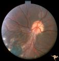 |
Neurofibromatosis-2 | Retinal tumor in NF-2 referred to as a CPERH (choroidal pigment epithelial retinal hamartoma). Patient, a 16 year old girl, had bilateral acoustic neurinomas. Pair with R1_F2b. Same eye. Anatomy: Optic disc; Retina. Pathology: Retinal hamartoma; Bilateral acoustic neurinoma. Disease/Diagnosis: Neuro... | Image |
| 303 |
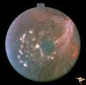 |
Neurofibromatosis-2 | Retinal tumor in NF-2 referred to as a CPERH (choroidal pigment epithelial retinal hamartoma). Patient, a 16 year old girl, had bilateral acoustic neurinomas. Pair with R1_F2a. Same eye. Anatomy: Optic disc; Retina. Pathology: Retinal hamartoma; Bilateral acoustic neurinoma. Disease/Diagnosis: Neuro... | Image |
| 304 |
 |
Neurofibromatosis-2 | This is the ocular fundus in a patient with NF-2 showing a preretinal membrane that extends from the temporal disc margin toward the macula. The optic disc shows low grade papilledema caused by one of the patient's acoustic neurinomas. The membrane has caused horizontal folds on the retinal surface.... | Image |
| 305 |
 |
P50 Chronic Papilledema with Subretinal Neo-Vascular Network | Chronic papilledema with subretinal neo-vascular network. Pseudotumor. Anatomy: Optic disc. Pathology: Papilledema. Disease/ Diagnosis: Chronic papilledema with sub-retinal neovascular network. | Image |
| 306 |
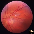 |
P52a Asymmetric Papilledema with Choroidal Folds | Left eye. Choroidal folds with no papilledema. Asymmetric papilledema with choroidal folds. Bilateral choroidal folds from elevated intracranial pressure. 52a. Anatomy: Optic disc. Pathology: Papilledema. Disease/ Diagnosis: Asymmetric - No papilledema with choroidal folds | Image |
| 307 |
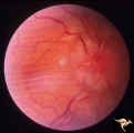 |
P52b Asymmetric Papilledema with Choroidal Folds | Right eye shows papilledema. Asymmetric papilledema with choroidal folds. Bilateral choroidal folds from elevated intracranial pressure. Anatomy: Optic disc. Pathology: Papilledema. Disease/ Diagnosis: Chronic papilledema with choroidal folds. | Image |
| 308 |
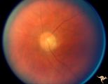 |
Paraneoplastic Retinopathy | Man with oat cell carcinoma of the lung with paraneoplastic retinopathy. Narrowed arterioles. Note absence of obvious retinal pigmentary degeneration. Cancer associated retinopathy syndrome (CAR Syndrome or Sawyer-Sellhorst Syndrome) (Ref: Sawyer, Sellhorst, Hoyt). Anatomy: Retina. Pathology: Oat c... | Image |
| 309 |
 |
Paraneoplastic Retinopathy | Man with oat cell carcinoma of the lung with paraneoplastic retinopathy. Narrowed arterioles. Note absence of obvious retinal pigmentary degeneration. Cancer associated retinopathy syndrome (CAR Syndrome or Sawyer-Sellhorst Syndrome) (Ref: Sawyer, Sellhorst, Hoyt). Anatomy: Retina. Pathology: Oat c... | Image |
| 310 |
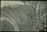 |
Pigment Epithelial Hamartoma of Optic Disc | Optic disc tumor discovered incidentally in a 32 year old Asian woman who had no complaints about visual function in her involved left eye. Fundus slide shows granular elevation of left disc obscurring major disc vessels. Some of the granules has a shiny crystalline appearance. Near the vessel entra... | Image |
| 311 |
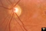 |
Pigmentary Retinopathy with Peripheral Neuropathy (Flecked Retinal Syndrome or Fundus Flavimaculatus) | Pigmentary retinopathy with peripheral neuropathy (Flecked Retinal Syndrome or Fundus Flavimaculatus) in a young woman. Anatomy: Retina. Pathology: Peripheral nerve degeneration. Disease/Diagnosis: Retinitis pigmentosa with hereditary peripheral degeneration. Clinical: Blindness. | Image |
| 312 |
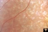 |
Pigmentary Retinopathy with Peripheral Neuropathy (Flecked Retinal Syndrome or Fundus Flavimaculatus) | Pigmentary retinopathy with peripheral neuropathy (Flecked Retinal Syndrome or Fundus Flavimaculatus) in a young woman. Anatomy: Retina. Pathology: Peripheral nerve degeneration. Disease/Diagnosis: Retinitis pigmentosa with hereditary peripheral degeneration. Clinical: Blindness. | Image |
| 313 |
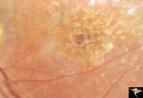 |
Pigmentary Retinopathy with Peripheral Neuropathy (Flecked Retinal Syndrome or Fundus Flavimaculatus) | Pigmentary retinopathy with peripheral neuropathy (Flecked Retinal Syndrome or Fundus Flavimaculatus) in a young woman. Anatomy: Retina. Pathology: Peripheral nerve degeneration. Disease/Diagnosis: Retinitis pigmentosa with hereditary peripheral degeneration. Clinical: Blindness. | Image |
| 314 |
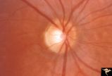 |
Pigmentary Retinopathy with Peripheral Neuropathy (Flecked Retinal Syndrome or Fundus Flavimaculatus) | Pigmentary retinopathy with peripheral neuropathy (Flecked Retinal Syndrome or Fundus Flavimaculatus) in a young woman. Anatomy: Retina. Pathology: Peripheral nerve degeneration. Disease/Diagnosis: Retinitis pigmentosa with hereditary peripheral degeneration. Clinical: Blindness. | Image |
| 315 |
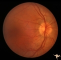 |
Post Papilledema | Right eye. Post Papilledema with minimal optic disc changes after treatment for temporal lobe glioma. Minimal optic disc haze. Optic disc. Pathology: Papilledema. Disease/Diagnosis: Post Papilledema due to temporal lobe glioma. | Image |
| 316 |
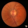 |
Post Papilledema | Left eye. Post Papilledema with minimal optic disc changes after treatment for temporal lobe glioma. Minimal optic disc haze. | Image |
| 317 |
 |
Post Papilledema, Secondary Optic Atrophy | Right eye. Post papilledema with chronic gliosis. arterial narrowing. ""high-water"" marks. Man. Anatomy: Optic disc. Pathology: Post papilledema. Disease/Diagnosis: Post papilledema with optic atrophy. | Image |
| 318 |
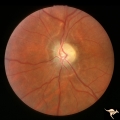 |
Post Papilledema, Secondary Optic Atrophy | Left eye. Post papilledema with chronic gliosis. arterial narrowing. "high-water" marks. Man. Anatomy: Optic disc. Pathology: Post papilledema. Disease/Diagnosis: Post papilledema with optic atrophy. | Image |
| 319 |
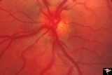 |
PP5b Crowded Disc | PP5a: left eye;PP5 b: left eye X 2 magnification; congenital disc blurring. Boy. Anatomy: Optic disc. Pathology: Normal variation of the optic disc. Disease/ Diagnosis: Normal variation of the optic disc. Congenital blurred disc. Clinical: Blurred disc margin. Beautiful example of difficult differen... | Image |
| 320 |
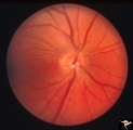 |
PP6a Crowded Disc with Glial Remnant | PP6a: 35 year old man. Right eye that has the glial remnant and blurred margins. PP6b: left eye. Anatomy: Optic disc. Pathology: Normal variation of the optic disc with glial remnant. Disease/ Diagnosis: Normal variation of the optic disc. Crowded disc with glial remnant. Clinical: Man referred for ... | Image |
| 321 |
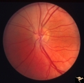 |
PP6b Crowded Disc with Glial Remnant | PP6a: right eye that has the glial remnant and blurred margins. PP6b: left eye. Anatomy: Optic disc. Pathology: Normal variation of the optic disc. Disease/ Diagnosis: Normal variation of the optic disc. Crowded disc. Clinical: Left eye is normal. | Image |
| 322 |
 |
PP7a Crowded disc | PP7a: right eye crowded disc with blurred margin. Note anomalous vascular pattern and glial tissue on the disc; PP7b- left disc is cupless disc and normal. 10 year old girl with gonadal dysgenesis and growth retardation. Anatomy: Optic disc. Pathology: Normal variation of the optic disc. Disease/ Di... | Image |
| 323 |
 |
PP8a Crowded Disc with Significant Nasal Disc Blurring | Congenital nasal disc blurring. Myopic eyes. Thai girl patient. One wonders about vitreal adherence to the disc. PP 8a right eye. Pair with left eye in PP8b. Anatomy: Optic disc. Pathology: Normal variation of the optic disc. Disease/ Diagnosis: Normal variation of the optic disc. Congenital blurre... | Image |
| 324 |
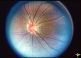 |
PP8b Crowded Disc with Significant Nasal Disc Blurring | Congenital nasal disc blurring. Myopic eyes. Thai girl patient. One wonders about vitreal adherence to the disc. PP 8b left eye. Pair with PP 8a right eye. Anatomy: Optic disc. Pathology: Normal variation of the optic disc. Disease/ Diagnosis: Normal variation of the optic disc. Congenital blurred d... | Image |
| 325 |
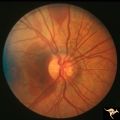 |
Pseudoxanthoma Elasticum (PXE) | Pseudoxanthoma elasticum (PXE) Right eye shows angiod streaks with associated hemorrhage. Patient was 25 year old man who developed a right sided carotid cavernous fistula. Anatomy: Retina. Pathology: Pseudoxanthoma elasticum (PXE). Disease/Diagnosis: PXE with angiod streaks with associated hemorrha... | Image |
