Best known for his world-renowned neuro-ophthalmology unit based at the University of California, San Francisco, William Hoyt, MD collected here more than 850 of his best images covering a wide range of disorders.
William F. Hoyt, MD, Professor Emeritus of Ophthalmology, Neurology and Neurosurgery, Department of Ophthalmology, University of California, San Francisco.
NOVEL: https://novel.utah.edu/
TO
| Title | Curriculum | Description | Subject | Collection | ||
|---|---|---|---|---|---|---|
| 51 |
 |
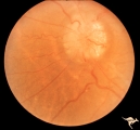
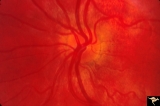
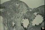
Vascular Complications of Drusen | PP34a: Right eye. Superior retinal vein drains into the choroid at 12:00. It has occluded between center of disc and 12:00. Note white ghost vessel. Note that other veins drain into the disc edge at 4:00. There is no evidence of a central retinal vein in the middle of the disc. PP34b: Visible drus... | Pseudopapilledema; Drusen Discs with Complications | Neuro-Ophthalmology Virtual Education Library: William F. Hoyt Neuro-Ophthalmology Collection: https://novel.utah.edu/Hoyt/ | |
| 52 |
 |
Vascular Complications of Drusen | PP34a: Right eye. Superior retinal vein drains into the choroid at 12:00. It has occluded between center of disc and 12:00. Note white ghost vessel. Note that other veins drain into the disc edge at 4:00. There is no evidence of a central retinal vein in the middle of the disc. PP34b: Visible drus... | Pseudopapilledema; Drusen Discs with Complications | Neuro-Ophthalmology Virtual Education Library: William F. Hoyt Neuro-Ophthalmology Collection: https://novel.utah.edu/Hoyt/ | |
| 53 |
 |
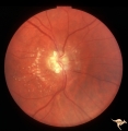
Chronic Papilledema with Pseudo Drusen | Chronic papilledema with pseudo drusen. Residual choroidal folds. Pseudo drusen. Anatomy: Optic disc. Pathology: Papilledema. Disease/Diagnosis: Chronic papilledema with pseudo drusen. | Papilledema; Complications | Neuro-Ophthalmology Virtual Education Library: William F. Hoyt Neuro-Ophthalmology Collection: https://novel.utah.edu/Hoyt/ | |
| 54 |
 |
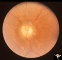
Visible Drusen with Visual Field Loss | Left eye visual field. Combine with PP25 a, b, & c. Anatomy: Optic disc. Pathology: Drusen of the optic disc. Disease/Diagnosis: Drusen of the optic disc. Clinical: Note marked constriction of visual field in all quadrants with central preservation of vision. | Pseudopapilledema; Exposed Drusen | Neuro-Ophthalmology Virtual Education Library: William F. Hoyt Neuro-Ophthalmology Collection: https://novel.utah.edu/Hoyt/ | |
| 55 |
 |
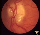
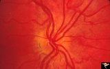
Venous Anomalies - Exit Anomalies | Intrapapillary drusen causing diversion of retinal venous blood into the disc edge at 12:00 and 4:00. Note the white ghost veins between the disc edge and the center of the disc. Anatomy: Optic disc. Pathology: Optic nerve drusen. Disease/Diagnosis: Complication of optic nerve drusen. Venous rerouti... | Vascular Disc Anomalies; Venous Anomalies; Exit Anomalies | Neuro-Ophthalmology Virtual Education Library: William F. Hoyt Neuro-Ophthalmology Collection: https://novel.utah.edu/Hoyt/ | |
| 56 |
 |
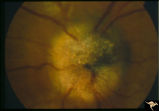
Chronic Papilledema with Pseudo Drusen | Left eye. Chronic papilledema with pseudo drusen due to cerebral pontine angle tumor. Anatomy: Optic disc. Pathology: Papilledema Disease/Diagnosis: Chronic papilledema with pseudo drusen. | Papilledema; Complications | Neuro-Ophthalmology Virtual Education Library: William F. Hoyt Neuro-Ophthalmology Collection: https://novel.utah.edu/Hoyt/ | |
| 57 |
 |
Chronic Papilledema with Pseudo Drusen | Right eye. Chronic papilledema with pseudo drusen due to cerebral pontine angle tumor. Anatomy: Optic disc Pathology: Papilledema Disease/Diagnosis: Chronic papilledema with pseudo drusen | Papilledema; Complications | Neuro-Ophthalmology Virtual Education Library: William F. Hoyt Neuro-Ophthalmology Collection: https://novel.utah.edu/Hoyt/ | |
| 58 |
 |
Chronic Papilledema with Pseudo Drusen | Right eye. Meningioma. Pseudo drusen from chronic papilledema. Woman. Anatomy: Optic disc Pathology: Papilledema Disease/Diagnosis: Chronic papilledema with pseudo drusen | Papilledema; Complications | Neuro-Ophthalmology Virtual Education Library: William F. Hoyt Neuro-Ophthalmology Collection: https://novel.utah.edu/Hoyt/ | |
| 59 |
 |
Chronic Papilledema with Pseudo Drusen | Left eye of 51 year old, 220 pound black woman. Pseudotumor cerebri, pseudo drusen, exudates. Anatomy: Optic disc. Pathology: Papilledema Disease/Diagnosis: Chronic papilledema with pseudo drusen | Papilledema; Complications | Neuro-Ophthalmology Virtual Education Library: William F. Hoyt Neuro-Ophthalmology Collection: https://novel.utah.edu/Hoyt/ | |
| 60 |
 |
Chronic Papilledema with Pseudo Drusen | Left eye. Meningioma. Pseudo drusen from chronic papilledema. The patient's meningioma had blinded her left eye and caused chronic elevated intracranial pressure. Woman. Anatomy: Optic disc Pathology: Papilledema Disease/Diagnosis: Chronic papilledema with pseudo drusen | Papilledema; Complications | Neuro-Ophthalmology Virtual Education Library: William F. Hoyt Neuro-Ophthalmology Collection: https://novel.utah.edu/Hoyt/ | |
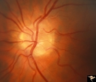
| 61 |
 |
Crowded Disc (Family) | curriculum_fellow; IC-C6biv-pseudo-disc-edema; KBDcongenitalblurreddisc; KBDdooncongenital;; KBDdcapseudocongenitalblurreddisc | Anomalous vasculature with congenital disc margin blurring. Note optic cup is absent. Pair with brother in PP1a & b. Mother has drusen of the optic disc in PP11aa & b. Sister has drusen in PP11c. Anatomy: Optic disc. Pathology: Normal variant. Cause of appearance is too many fibers entering into a s... | Pseudopapilledema; Congenital Blurred Disc | Neuro-Ophthalmology Virtual Education Library: William F. Hoyt Neuro-Ophthalmology Collection: https://novel.utah.edu/Hoyt/ |
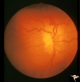
| 62 |
 |
Bilateral Crowded Discs | curriculum_fellow; KBDopticdiscanomalies; KBDcongenitalblurreddisc; KBDdooncongenital;; KBDdcapseudocongenitalblurreddisc | Left eye. Bilateral crowded discs with congenital blurring. Blurred disc margins are not from edema. Note optic cup is absent. Pair with right eye in PP_1a, and brother in PP_2. Mother has drusen of the optic disc in PP_11aa & b. Sister has drusen in PP_11c. Anatomy: Optic disc. Pathology: Normal va... | Pseudopapilledema; Congenital Blurred Disc; Optic Disc Anomalies | Neuro-Ophthalmology Virtual Education Library: William F. Hoyt Neuro-Ophthalmology Collection: https://novel.utah.edu/Hoyt/ |
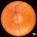
| 63 |
 |
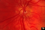
Bilateral Crowded Discs (Family) | curriculum_fellow; KBDopticdiscanomalies; KBDcongenitalblurreddisc; KBDdooncongenital;; KBDdcapseudocongenitalblurreddisc | Right eye. Bilateral crowded discs with congenital blurring. Blurred disc margins are not from edema. Note optic cup is absent. Pair with left eye in PP_1b, and brother in PP_2. Mother has drusen of the optic disc in PP_11a & b. Sister has drusen in PP_11c. Anatomy: Optic disc. Pathology: Normal var... | Pseudopapilledema; Congenital Blurred Disc; Optic Disc Anomalies | Neuro-Ophthalmology Virtual Education Library: William F. Hoyt Neuro-Ophthalmology Collection: https://novel.utah.edu/Hoyt/ |
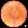
| 64 |
 |
IB201 Post Radiation Papillopathy | Right eye, 1982, Bizarre drusen like bodies on the pale atrophic disc. Note arteriolar narrowing. Note peripapillary circumferential retinal exudate. Anatomy: Optic disc. Pathology: Post radiation papillopathy. Disease/ Diagnosis: Post radiation papillopathy. Clinical: Blindness following radiation ... | Optic Disc Atrophy with Special Features; Focally Narrowed Retinal Arterioles | Neuro-Ophthalmology Virtual Education Library: William F. Hoyt Neuro-Ophthalmology Collection: https://novel.utah.edu/Hoyt/ | |
| 65 |
 |
F401 Pigment Epithelial Hamartoma of Optic Disc | Optic disc tumor discovered incidentally in a 32 year old Asian woman who had no complaints about visual function in her involved left eye. Fundus slide shows granular elevation of left disc obscurring major disc vessels. Some of the granules has a shiny crystalline appearance. Near the vessel entra... | Pigment Epithelium of Eye; Optic Disc | Neuro-Ophthalmology Virtual Education Library: William F. Hoyt Neuro-Ophthalmology Collection: https://novel.utah.edu/Hoyt/ | |
| 66 |
 |
F403 Pigment Epithelial Hamartoma of Optic Disc | Optic disc tumor discovered incidentally in a 32 year old Asian woman who had no complaints about visual function in her involved left eye. Fundus slide shows granular elevation of left disc obscuring major disc vessels. Some of the granules has a shiny crystalline appearance. Near the vessel entran... | Pigment Epithelium of Eye; Optic Disc | Neuro-Ophthalmology Virtual Education Library: William F. Hoyt Neuro-Ophthalmology Collection: https://novel.utah.edu/Hoyt/ | |
| 67 |
 |
Pigment Epithelial Hamartoma of Optic Disc | Optic disc tumor discovered incidentally in a 32 year old Asian woman who had no complaints about visual function in her involved left eye. Fundus slide shows granular elevation of left disc obscurring major disc vessels. Some of the granules has a shiny crystalline appearance. Near the vessel entra... | Pigment Epithelium of Eye; Optic Disc | Neuro-Ophthalmology Virtual Education Library: William F. Hoyt Neuro-Ophthalmology Collection: https://novel.utah.edu/Hoyt/ |