Best known for his world-renowned neuro-ophthalmology unit based at the University of California, San Francisco, William Hoyt, MD collected here more than 850 of his best images covering a wide range of disorders.
William F. Hoyt, MD, Professor Emeritus of Ophthalmology, Neurology and Neurosurgery, Department of Ophthalmology, University of California, San Francisco.
NOVEL: https://novel.utah.edu/
TO
| Title | Description | Type | ||
|---|---|---|---|---|
| 1 |
 |
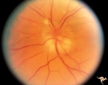
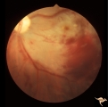
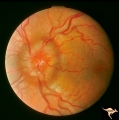
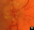
C110 Papillitis, Retrobulbar Neuritis | AIDs papillitis. Segmental. Note inflammatory focus on temporal side of disc. 29 year old homosexual male. Visual field shows huge blind spot. Anatomy: Optic disc. Pathology: Axoplasmic stasis due to inflammation. Disease/ Diagnosis: AIDS papillitis / AIDS Optic neuritis. Clinical: Visual symptoms d... | Image |
| 2 |
 |
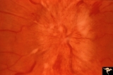
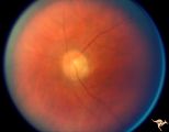
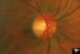
C111 Papillitis, Retrobulbar Neuritis | AIDS papillitis. Male. Anatomy: Optic disc. Pathology: Axoplasmic stasis due to inflammation. Disease/ Diagnosis: AIDS papillitis. Clinical: Visual symptoms. | Image |
| 3 |
 |
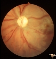
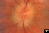
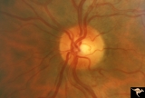
C402 Luetic Papillopathy (Gumma of the Optic Disc) | November 2001. Same eye as C4_01 after treatment with penicillin. Disc swelling went away and good visual function returned. Anatomy: Optic disc. Pathology: Axoplasmic stasis due to syphillitic infection. Disease/ Diagnosis: Luetic papillopathy (Syphillis). Clinical: Improving visual loss. | Image |
| 4 |
 |
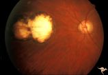
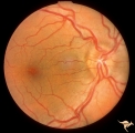
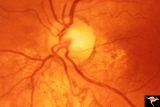
C401 Luetic Papillopathy (Gumma of the Optic Disc) | Diffuse optic disc swelling with tortuous capillary dilations indicating inflammatory cellular infiltration. October 2001. Same eye as C4_02. Anatomy: Optic disc. Pathology: Axoplasmic stasis due to syphillitic infection. Luetic papillopathy (Syphyllis). Clinical: Visual loss. | Image |
| 5 |
 |
H36 Segmental Hypoplasia, Retinal, Congenital Toxo | Left eye. Optic disc hypoplasia from congenital nasal retinal toxoplasma lesion. Chorioretinal scar. Anatomy: Optic disc, retina. Pathology: Hypoplasia secondary to retinal lesion. Disease/ Diagnosis: Segmental optic disc hypoplasia. | Image |
| 6 |
 |
Bilateral Papilledema with Pseudotumor Cerebri | Right eye. Mild bilateral papilledema in a 7 year old boy. Cause of swelling unknown. Growth failure treated with thyroid medication. Anatomy: Optic disc. Pathology: Bilateral papilledema. Disease/Diagnosis: Intracranial hypertension due to treatment of growth failure with thyroid medicaltion. Clini... | Image |
| 7 |
 |
Bilateral Papilledema with Pseudotumor Cerebri | Left eye. Mild bilateral papilledema in a 7 year old boy. Cause of swelling unknown. Growth failure treated with thyroid medication. Anatomy: Optic disc. Pathology: Bilateral papilledema. Disease/Diagnosis: Intracranial hypertension due to treatment of growth failure with thyroid medication. Clinica... | Image |
| 8 |
 |
G206 Purtchers Traumatic Retinopathy | Left eye. After auto accident in which the patient's chest was squeezed. Same eye as G2_07. Anatomy: Optic disc. Pathology: Varied peripapillary ischemic retinopathy. Disease/ Diagnosis: Purtchers traumatic retinopathy. | Image |
| 9 |
 |
G207 Purtchers Traumatic Retinopathy | Left eye. Large pre-retinal hemorrhage. Same eye as G2_06. Anatomy: Optic disc. Pathology: Varied peripapillary ischemic retinopathy. Disease/ Diagnosis: Purtchers traumatic retinopathy. | Image |
| 10 |
 |
Paraneoplastic Retinopathy | Man with oat cell carcinoma of the lung with paraneoplastic retinopathy. Narrowed arterioles. Note absence of obvious retinal pigmentary degeneration. Cancer associated retinopathy syndrome (CAR Syndrome or Sawyer-Sellhorst Syndrome) (Ref: Sawyer, Sellhorst, Hoyt). Anatomy: Retina. Pathology: Oat c... | Image |
| 11 |
 |
Paraneoplastic Retinopathy | Man with oat cell carcinoma of the lung with paraneoplastic retinopathy. Narrowed arterioles. Note absence of obvious retinal pigmentary degeneration. Cancer associated retinopathy syndrome (CAR Syndrome or Sawyer-Sellhorst Syndrome) (Ref: Sawyer, Sellhorst, Hoyt). Anatomy: Retina. Pathology: Oat c... | Image |
| 12 |
 |
Bilateral Papilledema with Pseudotumor Cerebri | Chronic appearance of swelling in right eye. 29 year old woman. Bilateral papilledema. Anatomy: Optic disc. Pathology: Bilateral papilledema. Disease/Diagnosis: Intracranial hypertension due to treatment of growth failure with thyroid medication. Clinical: symptoms: headache, signs: bilateral papill... | Image |
| 13 |
 |
Unilateral Papilledema | Right eye. Has no optic cup. Disc is flat. Anatomy: Optic disc. Pathology: Unilateral papilledema. Disease/Diagnosis: Idiopathic intracranial hypertension (pseudotumor cerebri). Clinical: Transient monocular blindness (transient visual obscurations, amaurosis fugax); headache, sixth nerve palsy, ele... | Image |
| 14 |
 |
Unilateral Papilledema | Left eye. This eye has papilledema. Anatomy: Optic disc. Pathology: Unilateral papilledema. Disease/Diagnosis: Idiopathic intracranial hypertension (pseudotumor cerebri). Clinical: Transient monocular blindness (transient visual obscurations, amaurosis fugax); headache, sixth nerve palsy, elevated i... | Image |
| 15 |
 |
Unilateral Papilledema | Unilateral papilledema in Pseudotumor cerebri. Right eye. Has no cup. Woman. Anatomy: Optic disc. Pathology: Unilateral papilledema. Disease/Diagnosis: Idiopathic intracranial hypertension, pseudotumor cerebri. Clinical: Woman, headache. | Image |
| 16 |
 |
Unilateral Papilledema | Left eye. Has chronic papilledema. Woman. Anatomy: Optic disc. Pathology: Unilateral papilledema. Disease/Diagnosis: Idiopathic intracranial hypertension, pseudotumor cerebri. Clinical: Woman, headache. | Image |
| 17 |
 |
Segmental Atrophy - Temporal | Segmental Atrophy - Temporal - Nutritional Amblyopia (alcohol) Discs show bilateral temporal pallor with hyperemia of the remaining disc tissue - Pair with IIA2_02b. 1971. Anatomy: Optic disc. Pathology: Optic atrophy. Disease/Diagnosis: Toxic optic atrophy from alcohol. Clinical: Central visual lo... | Image |
| 18 |
 |
Segmental Atrophy - Temporal | Segmental Atrophy - Temporal - Nutritional Amblyopia (alcohol) Discs show bilateral temporal pallor with hyperemia of the remaining disc tissue - Pair with IIA2_02a. 1971. Anatomy: Optic disc. Pathology: Optic atrophy. Disease/Diagnosis: Toxic optic atrophy from alcohol. Clinical: Central visual lo... | Image |
| 19 |
 |
Segmental Atrophy - Temporal | Segmental Atrophy - Temporal - Nutritional amblyopia (alcoholic). 1985. Left eye. Pair with IIA2_03a. Anatomy: Optic disc.. Pathology: Optic atrophy. Disease/Diagnosis: Toxic optic atrophy from alcohol. Clinical: Central visual loss. | Image |
| 20 |
 |
Segmental Atrophy - Hemianopic (Band) Atrophy from Eight Optic Tract Injury | Segmental Atrophy - Band atrophy from right optic tract injury. This eye has a nasal hemianopia. Its disc shows temporal pallor with an intact nasal nerve fiber layer. Pair with IIA2C_7b. Anatomy: Optic disc. Pathology: Right optic tract injury. Disease/Diagnosis: Homonymous hemioptic atrophy. Clini... | Image |
| 21 |
 |
Segmental Atrophy - Hemianopic (Band) Atrophy from Eight Optic Tract Injury | Segmental Atrophy - Band atrophy from right optic tract injury. Left eye. Has temporal hemianopia with band atrophy. Note loss of nasal nerve fiber layer. Four and a half months after injury from intracranial pressure catheter. Pair with IIA2C_7a. Anatomy: Optic disc. Pathology: Right optic tract in... | Image |
| 22 |
 |
Segmental Atrophy - Temporal | Segmental Atrophy - Temporal pallor - Nutritional amblyopia (alcoholic). 1985. Right eye. Pair with IIA2_03b. Anatomy: Optic disc. Pathology: Optic atrophy. Disease/Diagnosis: Toxic optic atrophy from alcohol. Clinical: Central visual loss. | Image |
| 23 |
 |
B103 Disc Swelling, Ischemic Papillopathies, AION | Ischemic swelling. 50 year old woman, 12 days after a viral illness. Nasal nerve fiber layer bundle visual field defect. Anatomy: Optic disc. Pathology: Axoplasmic stasis due to ischemia. Disease/ Diagnosis: AION. Clinical: Visual loss after viral illness. | Image |
| 24 |
 |
B102 Disc Swelling, Ischemic Papillopathies, AION | Ischemic swelling. March 2, 1978. Same patient as B1_03. Anatomy: Optic disc. Pathology: Axoplasmic stasis due to ischemia. Disease/ Diagnosis: AION. Clinical: Diabetic with optic disc swelling and visual loss. | Image |
| 25 |
 |
B203 Disc Swelling, Diabetic Papillopathy | Disc swelling in a diabetic woman. Recovered without visual loss. Right eye. Pair with B2_04. Anatomy: Optic disc. Pathology: Axoplasmic stasis due to ischemia. Disease/ Diagnosis: Diabetic papillopathy. Clinical: Visual loss with recovery. | Image |