Best known for his world-renowned neuro-ophthalmology unit based at the University of California, San Francisco, William Hoyt, MD collected here more than 850 of his best images covering a wide range of disorders.
William F. Hoyt, MD, Professor Emeritus of Ophthalmology, Neurology and Neurosurgery, Department of Ophthalmology, University of California, San Francisco.
NOVEL: https://novel.utah.edu/
TO
Filters: Collection: "ehsl_novel_wfh" Date: "1997"
1 - 25 of 7
| Title | Description | Type | ||
|---|---|---|---|---|
| 1 |
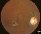 |
Cerebellar Macular Degenerative Disease | Ocular fundus shows prominent retinal degeneration in the region of the maculae, bilateral optic disc pallor with narrowed retinal arterioles. Interesting peripapillary halo of retinal pigment degeneration. Most consistent with Spinal Cerebellar Degeneration Type 7 (SCA-7). Anatomy: Retina. Patholog... | Image |
| 2 |
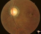 |
Cerebellar Macular Degenerative Disease | Ocular fundus shows prominent retinal degeneration in the region of the maculae, bilateral optic disc pallor with narrowed retinal arterioles. Interesting peripapillary halo of retinal pigment degeneration. Most consistent with Spinal Cerebellar Degeneration Type 7 (SCA-7). Anatomy: Retina. Patholog... | Image |
| 3 |
 |
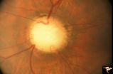
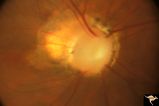
H03 Panhypoplasia | Extreme hypoplasia. Note absence of retinal nerve fiber layer. Left eye. Girl. Same patient as H_4. Anatomy: Optic disc. Pathology: Hypoplasia of the optic nerve. Disease/ Diagnosis: Hypoplasia. Clinical: Left eye. Girl. | Image |
| 4 |
 |
H04 Panhypoplasia | Right eye. Normal eye. Girl. Same patient as H_3. Anatomy: Optic disc. Pathology: Hypoplasia of the optic nerve. Disease/ Diagnosis: Hypoplasia. | Image |
| 5 |
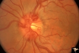 |
Venous Anomalies - Prepapillary Venous Convolutions (Congenital) | Prepapillary venous convolutions - congenital. 42 year old man. Incidental finding. Anatomy: Optic disc. Pathology: Prepapillary venous convolutions - congenital. Disease/Diagnosis: Prepapillary venous convolutions - congenital. Clinical: Asymptomatic. | Image |
| 6 |
 |
C29 Empty Disc | Left eye. Papilorenal Syndrome (PRS). Pair with C_30. Anatomy: Optic disc. | Image |
| 7 |
 |
C30 Empty Disc | Right eye. Papillorenal Syndrome (PRS). Pair with C_29. Anatomy: Optic disc. | Image |
1 - 25 of 7