Best known for his world-renowned neuro-ophthalmology unit based at the University of California, San Francisco, William Hoyt, MD collected here more than 850 of his best images covering a wide range of disorders.
William F. Hoyt, MD, Professor Emeritus of Ophthalmology, Neurology and Neurosurgery, Department of Ophthalmology, University of California, San Francisco.
NOVEL: https://novel.utah.edu/
TO
Filters: Collection: "ehsl_novel_wfh" Date: "1993"
1 - 25 of 12
| Title | Description | Type | ||
|---|---|---|---|---|
| 1 |
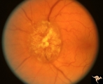 |
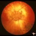
F104 Esthesio Neuroblastoma | Esthesioneuroblastoma. Tumor cells infiltrating the optic disc. 20/200 vision. Anatomy: Optic disc. Pathology: Esthesioneuroblastoma. Disease/ Diagnosis: Esthesioneuroblastoma. | Image |
| 2 |
 |
Retinal Signs of Atheromatous Embolization | Retinal signs of atheromatous embolization. Central retinal artery occlusion by soft atheromatous debris (mostly fibrin) causing blindness. Anatomy: Retina. Pathology: Carotid atheromatous disease. Disease/Diagnosis: Carotid atheromatous vascular disease. Clinical: Blindness. | Image |
| 3 |
 |
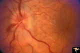
Vascular Disc Anomalies - Prepapillary Arterial Convolutions | Hemorrhage from prepapillary arterial convolutions has resolved. Abnormal vessels which were the source of the bleeding. 30 year old man. 3.5 months following hemorrhage. Same patient as V_10. Anatomy: Optic disc. Pathology: Congenital prepapillary arterial convolutions with pre-retinal hemorrhage.... | Image |
| 4 |
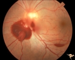 |
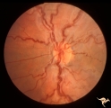
Vascular Disc Anomalies - Prepapillary Arterial Convolutions | Hemorrhage from prepapillary arterial convolutions. Note convolutions are inferior. 30 year old man. Same patient as V_11. Anatomy: Optic disc. Pathology: Congenital prepapillary arterial convolutions with pre-retinal hemorrhage. Disease/Diagnosis: Congenital arterial vascular anomaly. Clinical: Ac... | Image |
| 5 |
 |
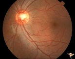
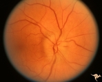
Vascular Disc Anomalies - Prepapillary Arterial Loop | Small central prepapillary arterial loop. 30 year old woman. Anatomy: Optic disc. Pathology: Congenital prepapillary arterial loop. Disease/Diagnosis: Congenital prepapillary arterial loop. Clinical: Asymptomatic. | Image |
| 6 |
 |
A101 Disc Swelling due to Intraocular Hypotension | Ocular hypotension following lens replacement surgery. Retinal/macular folds. Anatomy: Optic disc. Pathology: Disc edema. Disease/ Diagnosis: Intraocular hypotension. Clinical: Low intraocular pressure or intraocular hypotension. | Image |
| 7 |
 |
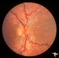
Vascular Disc Anomalies - Prepapillary Arterial Convolutions | Prepapillary arterial convolutions. Incidental finding in patient being treated for acute myelogenous leukemia. Note hemorrhage at about 4:00 off the disc related to the leukemia. Arterial loops are not related to leukemia. Anatomy: Optic disc. Pathology: Congenital prepapillary arterial convolution... | Image |
| 8 |
 |
Venous Anomalies - Congenital Venous Tortuosity | Congenital venous tortuosity. Left eye. 9 year old boy. Same patient as V_51. Anatomy: Optic disc. Pathology: Congenital venous tortuosity. Disease/Diagnosis: Congenital venous tortuosity. Clinical: Asymptomatic. | Image |
| 9 |
 |
Venous Anomalies - Congenital Venous Tortuosity | Congenital venous tortuosity. Right eye. 9 year old boy. Same patient as V_52. Anatomy: Optic disc. Pathology: Congenital venous tortuosity. Disease/Diagnosis: Congenital venous tortuosity. Clinical: Asymptomatic. | Image |
| 10 |
 |
C15 Morning Glory Disc | "Morning Glory" disc. Note tapering edge pointing to patient's transphenoidal encephalocele. Reference: Brodsky MC, Hoyt WF, Hoyt CS, Miller NR, Lam BL. Atypical retinochoroidal coloboma in patients with dysplastic optic discs and transphenoidal encephalocele Arch Ophthalmol. 1995 May;113(5):624-8.... | Image |
| 11 |
 |
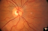
B111 Disc Swelling, Ischemic Papillopathies, AION | Acute AION. Anatomy: Optic disc. Pathology: Axoplasmic stasis due to ischemia. Disease/ Diagnosis: AION. Clinical: Visual loss. | Image |
| 12 |
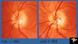 |
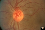
Von Hippel Lindau Disease | Von Hippel Lindau lesion on optic disc showing minimal increase in size over three year interval. Anatomy: Optic disc. Pathology: Hemangioblastoma. Disease/Diagnosis: Von Hippel Lindau disease. Clinical: Patient other eye was removed for hemangioblastoma. He has numerous hemangioblastomas of his spi... | Image |
1 - 25 of 12