Best known for his world-renowned neuro-ophthalmology unit based at the University of California, San Francisco, William Hoyt, MD collected here more than 850 of his best images covering a wide range of disorders.
William F. Hoyt, MD, Professor Emeritus of Ophthalmology, Neurology and Neurosurgery, Department of Ophthalmology, University of California, San Francisco.
NOVEL: https://novel.utah.edu/
TO
1 - 25 of 18
| Title | Description | Type | ||
|---|---|---|---|---|
| 1 |
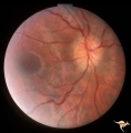 |
B112 Disc Swelling, Ischemic Papillopathies, AION | Arterioles are narrowing in resolution phase from AION. Patient had a superior altitudinal visual field defect. 20 year old man. Anatomy: Optic disc. Pathology: Axoplasmic stasis due to ischemia. Disease/ Diagnosis: AION. Clinical: Visual loss. | Image |
| 2 |
 |
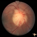
B114 Disc Swelling, Ischemic Papillopathies, AION | AION in a disc with an optic cup. Extraordinary exception with AION. Note ischemic vascular changes in disc surface. Anatomy: Optic disc. Pathology: Axoplasmic stasis due to ischemia. Disease/ Diagnosis: AION. Clinical: Visual loss. | Image |
| 3 |
 |
C07 Pits of the Optic Disc | Left eye. Temporal pit. Man. Anatomy: Optic disc. | Image |
| 4 |
 |
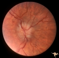
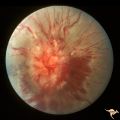
C304 Nodular Papillopathies (Sarcoid) | Lumpy nodular disc infiltration from sarcoid. Anatomy: Optic disc. Pathology: Axoplasmic stasis due to sarcoid infiltration. Disease/ Diagnosis: Sarcoid papillopathy. Clinical: Unknown? | Image |
| 5 |
 |
F102 Myeloblastic Leukemia | Myeloblastic leukemia. Left eye. Pair with F1_03. Anatomy: Optic disc. Pathology: Neoplastic (metastatic) papillopathy. Disease/ Diagnosis: Myeloblastic leukemia. | Image |
| 6 |
 |
F103 Myeloblastic Leukemia | Myeloblastic leukemia. Right eye. Pair with F1_02. Anatomy: Optic disc. Pathology: Neoplastic (metastatic) papillopathy. Disease/ Diagnosis: Myeloblastic leukemia. | Image |
| 7 |
 |
F109 T-Cell Leukemia Infiltrate | T-Cell leukemia infiltrate. 14 year old boy with T-Cell leukemia infiltrating the disc. Anatomy: Optic disc. Pathology: T-Cell leukemia. Disease/ Diagnosis: Neoplastic (metastatic) papillopathy | Image |
| 8 |
 |
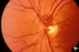
G102 Evulsion | Partial evulsion of the left optic nerve. Anatomy: Optic disc. Pathology: Optic nerve has been evulsed. Disease/ Diagnosis: Evulsion of the optic disc. | Image |
| 9 |
 |
H101 Occipital Hemianoptic Hypoplasia | Right eye. Same patient as H_102. Anatomy: Optic disc. Pathology: Occipital hemianoptic hypoplasia. Disease/ Diagnosis: Congenital defect of the occipital lobe. | Image |
| 10 |
 |
H102 Occipital Hemianoptic Hypoplasia | Left eye. Trans-synaptic band atrophy. Left homonymous hemianopia from right occipital porencephaly. Loss of nasal nerve fibers. Same patient as H_101. Anatomy: Optic disc. Pathology: Occipital hemianoptic hypoplasia. Disease/ Diagnosis: Congenital defect of the occipital lobe. | Image |
| 11 |
 |
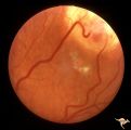
Sturge Weber Syndrome (Encephalotrigeminal Angiomatosis) | Sturge Weber Syndrome (Encephalotrigeminal angiomatosis) with retinal evidence of central retinal vein occlusion. Anatomy: Retina. Pathology: Diffuse choroidal hemangioma; Glaucoma. Disease/Diagnosis: Sturge Weber Syndrome. Clinical: Port wine hemangioma of the face. | Image |
| 12 |
 |
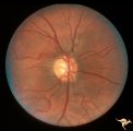
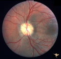
Vascular Disc Anomalies - Prepapillary Arterial Convolutions | Prepapillary arterial convolutions. 40 year old woman. Anatomy: Optic disc. Pathology: Congenital prepapillary arterial convolutions. Disease/Diagnosis: Congenital arterial vascular anomaly. Clinical: Asymptomatic. | Image |
| 13 |
 |
Visible Drusen with Visual Field Loss | Right eye visual field combine with PP25a, b, & d. Anatomy: Optic disc. Pathology: Drusen of the optic disc. Disease/Diagnosis: Drusen of the optic disc. Clinical: Drusen disc with severe visual field defect. note the nasal visual field loss and the arcuate bundle defects. Central vision was 20/20. | Image |
| 14 |
 |
Visible Drusen with Visual Field Loss | Left eye visual field. Combine with PP25 a, b, & c. Anatomy: Optic disc. Pathology: Drusen of the optic disc. Disease/Diagnosis: Drusen of the optic disc. Clinical: Note marked constriction of visual field in all quadrants with central preservation of vision. | Image |
| 15 |
 |
Visible Drusen with Visual Field Loss | PP25a: Left eye: Severe visual field defect. PP25b: right eye with exposed drusen and field loss: visual field defects; PP25c: right eye visual field PP25d: left eye visual field. Anatomy: Optic disc. Pathology: Drusen of the optic disc. Disease/Diagnosis: Drusen of the optic disc. Clinical: Dr... | Image |
| 16 |
 |
Visible Drusen with Visual Field Loss | PP25b right eye with drusen and severe visual field loss. Match with PP25a, c & d. Anatomy: Optic disc. Pathology: Drusen of the optic disc. Disease/Diagnosis: Drusen of the optic disc. Clinical: Drusen disc with servere visual field loss. | Image |
| 17 |
 |
Von Hippel Lindau Disease (Retinal Hemangioblastoma) | Von Hippel Lindau Disease (Retinal Hemangioblastoma); Small hemangioblastoma on the disc margin at 10:00. Large peripheral hemangioblastoma out of view to the top right seen on R1_C2b. Anatomy: Optic disc. Pathology: Hemangioblastoma. Disease/Diagnosis: Von Hippel Lindau disease. Clinical: No visual... | Image |
| 18 |
 |
Von Hippel Lindau Disease (Retinal Hemangioblastoma) | Von Hippel Lindau Disease (Retinal Hemangioblastoma); Eye with large peripheral hemangioblastoma. Note particularly the large draining vein. Pair with R1_C2a. Anatomy: Optic disc. Pathology: Hemangioblastoma. Disease/Diagnosis: Von Hippel Lindau disease. Clinical: No visual symptoms. | Image |
1 - 25 of 18