Collection of materials relating to neuro-ophthalmology as part of the Neuro-Ophthalmology Virtual Education Library.
NOVEL: https://novel.utah.edu/
TO
1 - 200 of 97
| Title | Creator | Description | Subject | ||
|---|---|---|---|---|---|
| 1 |
 |
Introduction to the NANOS Neuro-Ophthalmology Techniques of Examination (NOTE) | Karl C. Golnik, MD | An introduction to the NANOS Neuro-Ophthalmology Techniques of Examination (NOTE) | Examination; Eye Exam |
| 2 |
 |
Introduction to the Neurological Examination in NANOS NOTE | Padmaja Sudhakar, MD | An introduction to the neurological examination. | Neurology; Neurological Examination |
| 3 |
 |
Introduction to Examination of Eye Movements and Alignment in NANOS NOTE | Jason H. Peragallo, MD | Introduction to Examination of Eye Movements and Alignment | Eye Movement; Alignment; Examination |
| 4 |
 |
Introduction to Diagnostic Tests in NANOS NOTE | Amanda D. Henderson, MD | An introduction to Diagnostic Tests | Diagnostic Testing |
| 5 |
 |
Introduction to Examination of the Pupil in NANOS NOTE | Clare Fraser, MBBS, MMed | Introduction to Examination of the Pupil | Pupil; Examination |
| 6 |
 |
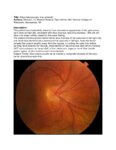
Introduction to Funduscopic Examination in NANOS NOTE | Rahul Sharma, MD, MPH | An introduction to Funduscopic Examination | Funduscopy; Examination |
| 7 |
 |
Introduction to Examination of the Orbit and the Extraocular Structures in NANOS NOTE | Julie Falardeau, MD | Introduction to Examination of the Orbit and the Extraocular Structures | Orbit; Anatomy |
| 8 |
 |
Internuclear Ophthalmoplegia | Wael A. Alsakran, MD; Valérie Biousse, MD | A slideshow describing INO; includes a video clip of saccadic delay. | INO |
| 9 |
 |
Introduction to Evaluation in Special Situations in NANOS NOTE | John Pula, MD | An introduction to NOTE sections on Examination of the Comatose Patient and Examination of the Pediatric Patient. | Patient Examination; Pediatric Patient; Examination |
| 10 |
 |
Botulinum Toxin and Migraine | Benjamin Frishberg, MD, FAAN, FNANOS | A video describing the use of botulinum toxin (Botox) for the treatment of migraine. | Botox; Botulinum Toxin; Migraine |
| 11 |
 |
Cataract Surgery | Laura L. Hanson, MD | A narrated slide presentation on the basics of cataract surgery. | Cataracts; Surgical Procedures |
| 12 |
 |
Optic Chiasm | Yesha Shah, BSA, BBA; Amanda Henderson, MD | Overview of the anatomy of the optic chiasm. | Optic Chiasm; Anatomy |
| 13 |
 |
Benign Essential Blepharospasm and Hemifacial Spasm | Celia Chen, MBBS, PhD, FRANZCO | Narrated lecture on treatment of benign essential blepharospasm and hemifacial spasm. | Benign Essential Blepharospasm; Hemifacial Spasm |
| 14 |
 |
Complications of Strabismus Surgery and Botox | W. Walker Motley, MD | A narrated video slideshow outlining complications associated with strabismus surgery. | Strabismus; Surgery; Surgical Complications; Botox |
| 15 |
 |
Inferior Orbital Fissure | Yesha Shah, BSA, BBA; Amanda Henderson, MD | Narrated lecture describing the inferior orbital fissure. | Inferior Orbital Fissure |
| 16 |
 |
Practice Based Learning and Improvement (PBLI) | Karl C. Golnik, MD, MEd | Video describing methods and best practices of Practice Based Learning and Improvement (PBLI). | Practice Based Learning and Improvement (PBLI) |
| 17 |
 |
Professionalism and Communication Skills | Karl C. Golnik, MD, MEd | Lecture covering professionalism and communication skills. | Professionalism; Communication Skills |
| 18 |
 |
Principles of Strabismus Surgery | Michael B. Yang, MD | A video demonstrating a medial rectus recession. | Strabismus; Surgery; Surgery Demonstrations |
| 19 |
 |
Anatomy of the Oculomotor Nerve (CN III) | Lucas E. Morgan, MS4; Nicholas A. Koontz, MD; Devin D. Mackay, MD | A detailed overview of the anatomic course of CN III, including a detailed pathway description and labeled MRI images, gross anatomy pictures, and structural models. | CN III; Third Cranial Nerve; Oculomotor Nerve; Anatomy; MRI |
| 20 |
 |
Visual Maturation | Yesha Shah, BSA, BBA; Amanda Henderson, MD | Video lecture covering visual maturation. | Visual Maturation; Foveal Development; Vision in Infants |
| 21 |
 |
Principles of Glaucoma Surgery | Aubrey Tirpack, MD | A video outlining the principles of glaucoma surgery for neuro-ophthalmologists. | Glaucoma; Glaucoma Surgery; Principles of Surgery |
| 22 |
 |
Common Patterns of Visual Field Defects | Sean Gratton, MD; Sarah Lam, 6th year BA/MD | Lecture covering common visual field defects, including those of the retina, optic nerve, chiasm, and retrochiasmal. | Visual Field Defects |
| 23 |
 |
Cotton Wool Spots: The Basics | Arnav Gupta, BHSc; Rahul Sharma, MD, MPH | A presentation describing cotton wool spots, an abnormal finding on funduscopic exam of the retina of the eye. | Cotton Wool Spots; Retina |
| 24 |
 |
Disc Cupping: The Basics | Arnav Gupta, BHSc; Rahul Sharma, MD, MPH | A presentation describing optic disc cupping, due to damage of optic nerve fibres. | Disc Cupping; Optic Disc |
| 25 |
 |
Retinal Detachment: The Basics | Arnav Gupta, BHSc; Rahul Sharma, MD, MPH | A presentation describing retinal detachment. | Retinal Detachment |
| 26 |
 |
Retinal Exudate: The Basics | Arnav Gupta, BHSc; Rahul Sharma, MD, MPH | A presentation describing retinal exudate. | Retinal Exudate |
| 27 |
 |
Retinal Hemorrhage: The Basics | Arnav Gupta, BHSc; Rahul Sharma, MD, MPH | A presentation describing retinal hemorrhage. | Retinal Hemorrhage |
| 28 |
 |
Introduction to the Evaluation of Visual Function in NANOS NOTE | Sean Gratton, MD | Introduction to the Evaluation of Visual Function in NANOS NOTE | Visual Function; Examination |
| 29 |
 |
Situs Inversus Optic Disc Anomaly | Michael Hii, Medical Student; Ryan Walsh, MD | This patient was incidentally-noted to have anomalous appearance of the optic discs, right more so than left, consistent with situs inversus optic disc anomaly. She did not have any visual deficits related to this exam finding. ; The patient's fundus photos demonstrate situs inversus of the optic ... | Situs Inversus Optic Disc Anomaly |
| 30 |
 |
Age-related Macular Degeneration: The Basics | Arnav Gupta, BHSc; Rahul Sharma, MD, MPH | A presentation covering age-related macular degeneration ("ARMD" or "AMD"), an acquired, progressive, chronic, degenerative disease of the retina. | Macular Degeneration |
| 31 |
 |
Temporal Artery Biopsy Procedure | Nooran Badeeb; Danah Albreiki | Temporal artery biopsy is a procedure that is done in a patient with suspicion of GCA (Giant cell Arteritis), and some of the clinical manifestations that prompts us to suspect the diagnosis in patients older than 50 years old are: 1. GCA symptoms e.g. new onset headache. 2 . Visual symptoms: - Visi... | Temporal Artery Biopsy; GCA; Temporal Arteritis |
| 32 |
 |
An Introduction to CT and MRI in Neuro-Imaging | Michael Carper, MD | A brief lecture covering basic neuro-imaging, including computed tomography (CT) and magnetic resonance imaging (MRI). | Computed Tomography (CT); Magnetic Resonance Imaging (MRI); Neuro-imaging |
| 33 |
 |
Progressive Supranuclear Palsy (PSP) | Molly Cincotta, MD; Ali G. Hamedani, MD, MHS | Objectives:; To provide an overview of PSP and its pathophysiology;; To present typical clinical features of the disease with a focus on ocular findings;; To provide a template for work up, diagnosis and treatment; ; To demonstrate typical eye movement abnormalities seen in PSP | Progressive Supranuclear Palsy (PSP) |
| 34 |
 |
Computed Tomography (CT): Principles, Technique, and Neuro-ophthalmic Applications | Alex Fraser, MD | Presentation covering Computed Tomography principles, adverse effects, comparison vs. MRI, and assorted examples of neuro-ophthalmic interest. | Computed Tomography (CT) |
| 35 |
 |
Examining the Pediatric Patient for Non-Neuro-ophthalmologists | John Pula, MD | Ten need-to-know pearls for examining the pediatric patient, for non-neuro-ophthalmologists. | Pediatric Patient Exam |
| 36 |
 |
Examining the Comatose Patient for Non-Neuro-ophthalmologists | John Pula, MD | Seven need-to-know pearls for examining the pediatric patient, for non-neuro-ophthalmologists. | Comatose Patient Exam |
| 37 |
 |
Visual Fields Part 2: Interpreting The Test Results | Jonathan Trobe, MD | Discussion of interpreting the results of visual field testing. | Visual Fields |
| 38 |
 |
Visual Fields Part 1: Performing The Tests | Jonathan Trobe, MD | Discussion and demonstration of visual field testing. | Visual Fields |
| 39 |
 |
Coordination Exam: Abnormal Examples: Heel-to-shin (x2) (includes Spanish audio & captions) | Paul D. Larsen, MD | The patient with ataxia of the lower extremity will have difficulty placing the heel on the knee with a side-to-side irregular over- and undershooting as the heel is advanced down the shin. Dysmetria on heel-to-shin can be seen in midline ataxia syndromes as well as cerebellar hemisphere disease so ... | Coordination Examination; Heel-shin Test |
| 40 |
 |
Coordination Exam: Normal Exam: Finger-to-nose (includes Spanish audio & captions) | Paul D. Larsen, MD | The patient moves her pointer finger from her nose to the examiner's finger as the examiner moves his finger to new positions and tests accuracy at the furthest outreach of the arm. NeuroLogic Exam has been supported by a grant from the Slice of Life Development Fund at the University of Utah, the D... | Coordination Examination; Finger-to-nose Test |
| 41 |
 |
Coordination Exam: Abnormal Examples: Finger-to-nose (x2) (includes Spanish audio & captions) | Paul D. Larsen, MD | The patient places her heel on the opposite knee then runs the heel down the shin to the ankle and back to the knee in a smooth coordinated fashion. NeuroLogic Exam has been supported by a grant from the Slice of Life Development Fund at the University of Utah, the Department of Pediatrics and the O... | Coordination Examination; Finger-to-nose Test |
| 42 |
 |
Coordination Exam: Normal Exam: Heel-to-shin (includes Spanish audio & captions) | Paul D. Larsen, MD | The patient places her heel on the opposite knee then runs the heel down the shin to the ankle and back to the knee in a smooth coordinated fashion. NeuroLogic Exam has been supported by a grant from the Slice of Life Development Fund at the University of Utah, the Department of Pediatrics and the O... | Coordination Examination; Heel-shin Test |
| 43 |
 |
Posterior Cortical Atrophy | Natali V. Baner, MD; Ali G. Hamedani, MD, MHS | PowerPoint providing an overview of the definition, clinical presentation and treatment of posterior cortical atrophy | Posterior Cortical Atrophy |
| 44 |
 |
Dementia: Overview and Classification | Molly Cincotta, MD; Whitley Aamodt, MD; Ali G. Hamedani, MD, MHS | PowerPoint providing a broad overview of dementia, including definition, clinical findings, work up, diagnosis, classification, and management. | Dementia |
| 45 |
 |
Multiple System Atrophy: Overview and Neuro-ophthalmologic Features | Pavan Vaswani, MD, PhD, Movement Disorders Fellow; Ali G. Hamedani, MD, MHS | Objectives: Know the key pathologic features of Multiple System Atrophy; Recognize the clinical presentation, including neuro-ophthalmologic features; Understand the symptomatic therapies and prognosis | Multiple System Atrophy |
| 46 |
 |
Dementia with Lewy Bodies: Overview and Neuro-ophthalmologic features | Pavan Vaswani, MD, PhD; Ali G. Hamedani, MD, MHS | Objectives: Recognize the difference between Dementia with Lewy Bodies and Parkinson disease dementia; Recognize the clinical presentation of DLB and differentiating features from Alzheimer disease dementia; Understand the symptomatic therapies and prognosis | Dementia; Lewy Bodies |
| 47 |
 |
Vascular Dementia | Whitley Aamodt, MD; Ali G. Hamedani, MD, MHS | PowerPoint providing an overview of vascular dementia, including the pathophysiology, clinical symptoms, diagnosis, and management. | Vascular Dementia |
| 48 |
 |
Tolosa Hunt Syndrome | Sahil Aggarwal, MD; Jason Liss, MD | Presentation covering an overview of Tolosa Hunt Syndrome. | Tolosa Hunt Syndrome |
| 49 |
 |
Amyotrophic Lateral Sclerosis (ALS) | Natali V. Baner, MD; Ali G. Hamedani, MD, MHS | PowerPoint providing an overview of the definition, clinical presentation and treatment of amyotrophic lateral sclerosis (ALS). | Amyotrophic Lateral Sclerosis (ALS) |
| 50 |
 |
Frontotemporal Dementia: Overview and Neuro-ophthalmologic Features | Pavan Vaswani, MD, PhD; Ali G. Hamedani, MD, MHS | Objectives: Understand the diagnostic criteria for the frontotemporal dementias; Differentiate behavioral variant FTD and the common variants of primary progressive aphasia; Recognize neuro-ophthalmologic and imaging features seen in FTD syndromes | Frontotemporal Dementia |
| 51 |
 |
Medicolegal and Ethical Considerations in Ophthalmology | M. Tariq Bhatti, MD | Slideshow describing topic. | Ethics; Legal |
| 52 |
 |
Maintenance of Certification Basics: American Board of Psychiatry and Neurology | Sean Gratton, MD | Video lecture covering certification basics. | Credentialing |
| 53 |
 |
New Evaluation and Management Rules for 2021 | Sean Gratton, MD | Overview of the coding rule changes implemented on January 1, 2021. | Coding Rules |
| 54 |
 |
Crafting a Grant Proposal for Research | Silvia Sörensen, PhD | Lecture describing the process of writing a grant for research. | Grant Writing |
| 55 |
 |
Grant Pieces - Research Grants | Silvia Sörensen, PhD | Lecture describing the parts of a research grant, including using human subjects. | Grant Writing |
| 56 |
 |
Diagnostic Error of Neuro-ophthalmologic Conditions: State of the Science | Leanne Stunkel, MD; David E. Newman-Toker, MD, PhD; Nancy J. Newman, MD; Valérie Biousse, MD | Diagnostic error is prevalent and costly, occurring in up to 15% of US medical encounters and affecting up to 5% of the US population. One-third of malpractice payments are related to diagnostic error. A complex and specialized diagnostic process makes neuro-ophthalmologic conditions particularly vu... | Diagnostic Errors |
| 57 |
 |
Protecting Human Subjects in Biomedical Research | Lisa R. Latchney, MS, CCRC | PowerPoint discussion of the history and development of ethics regulations in health research. | Ethical Issues in Research; Consent |
| 58 |
 |
Manuscripts: You Can Write These! | Elaine Smolock, PhD | Overview of writing techniques and parts of the manuscript, basic approach to writing results sections, what makes a good introduction, crafting a meaningful discussion, abstract and title suggestions, and how to get your editor's attention. | Writing Techniques |
| 59 |
 |
Radiation Optic Neuropathy | Neil R. Miller, MD, FACS | Overview of Radiation Optic Neuropathy (RON). | Radiation Optic Neuropathy; RON |
| 60 |
 |
Night Wolf | Mehdi Tavakoli, MD; Byron Lam, MD | A case presentation on radiation optic neurology. | Radiation Optic Neuropathy |
| 61 |
 |
Acute Optic Neuritis | Neil R. Miller, MD, FACS | Overview of acute optic neuritis. | Optic Neuritis |
| 62 |
 |
Afferent Visual Pathway Disorders: Typical vs Atypical Optic Neuritis | Carmen Chan, FRCP, FRCOphth, FRCSEd(Ophth), FHKAM(Ophthalmology) | Discussion of typical vs atypical optic neuritis. | Optic Neuritis |
| 63 |
 |
Superior Segmental Optic Disc Hypoplasia (SSOH) "Topless Disc Syndrome" | Sparsh Jain, Medical Student; Ryan Walsh, MD | This is a case of superior segmental optic disc hypoplasia that was found incidentally after a screening visual field test revealed an asymptomatic inferior field defect in the left eye. The patient has a unilateral SSOH in the left eye. | Superior Segmental Optic Disc Hypoplasia (SSOH) |
| 64 |
 |
Bergmeister Papilla | Sumayya Almarzouqi, MD | A brief overview of Bergmeister papilla, a rare congenital disc anomaly. It arises from the center of the optic disc consists of a small tuft of fibrous tissue and represents a remnant of the fetal hyaloid artery. | Bergmeister Papilla |
| 65 |
 |
Modern Imaging of Optic Disc Drusen | Meagan Seay, DO | This is a short powerpoint describing imaging techniques (specifically OCT-EDI, fundus autofluorescence, and B-scan ultrasonography) for optic disc drusen. Examples of these techniques are included. | Optic Disc Drusen; Imaging; OCT-EDI; Fundus Autofluorescence; B-scan Ultrasonography |
| 66 |
 |
Suprasellar Meningioma | Sumayya Almarzouqi, MD | Description of a case of suprasellar or sellar mass causeing chiasmal compression. | Suprasellar Meningioma |
| 67 |
 |
Bony Anatomy of the Orbit | Aakash Patel; Amanda Henderson, MD | Narrated presentation on the bony anatomy of the orbit. | Bony Anatomy; Orbit |
| 68 |
 |
Physiology of the Uvea | Aakash Patel; Amanda Henderson, MD | Overview of the physiology of the uvea. | Uvea; Physiology |
| 69 |
 |
Ocular Surface, Cornea, & Lens | Sari Yordi, MD | Video lecture on the anatomy of the ocular surface, cornea, and lens. | Ocular Surface; Cornea; Lens |
| 70 |
 |
Lacrimal Pathways: Anatomy and Physiology | Sari Yordi, MD | Video lecture covering anatomy and physiology of the lacrimal pathways. | Lacrimal Pathways |
| 71 |
 |
Aqueous and Vitreous Humor | Sari Yordi, MD | Narrated lecture on the aqueous and vitreous humor. | Aqueous; Vitreous Humor |
| 72 |
 |
Physiology of Intraocular Pressure | Aakash Patel; Amanda Henderson, MD | Overview of the physiology of intraocular pressure. | Intraocular Pressure; Physiology |
| 73 |
 |
Arteriovenous Malformation | Justin Gibson, MD; Charles Prestigiacomo, MD | A diagnostic cerebroangiogram performed on a patient who presented with worst headache of life, found to have a Fisher Grade 3 subarachnoid hemorrhage. | Angiogram; Arteriovenous Malformation; AVM |
| 74 |
 |
Large Vessel Occlusion | Justin Gibson, MD; Charles Prestigiacomo, MD | Example of a diagnostic cerebroangiogram performed on a patient undergoing a stroke. | Angiogram; Stroke |
| 75 |
 |
Cerebral Aneurysm | Justin Gibson, MD; Charles Prestigiacomo, MD | Cerebral angiogram of a patient with an arteriovenous malformation, or AVM. | Angiogram; Cerebral Aneurysm |
| 76 |
 |
Normal Angiogram | Justin Gibson, MD; Charles Prestigiacomo, MD | Example of a normal diagnostic cerebroangiogram. | Angiogram |
| 77 |
 |
Anaesthesia for Eye Surgery and Associated Complications | Julie Smith, MBBS, FANZCA | Lecture covering commonly performed eye surgery and anaesthetic techniques. | Eye Surgery; Anesthesia |
| 78 |
 |
Neuroablative Procedures | Benjamin Jonker, MB BS, MMed(Clin Epi), FRACS | Video lecture covering neuro-ablative procedures that are relevant to neuro-ophthalmologists. | Ablative Procedures |
| 79 |
 |
Anaesthesia for Eye Surgery and Associated Complications Slides | Julie Smith, MBBS, FANZCA | Lecture covering commonly performed eye surgery and anaesthetic techniques. | Eye Surgery; Anesthesia |
| 80 |
 |
Embryology of the Eye | Yesha Shah, BSA, BBA; Amanda Henderson, MD | Video lecture covering the embryology of the eye. | Embryology; Eye |
| 81 |
 |
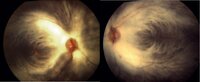
Myelinated Retinal Nerve Fibers | Scott N. Grossman, MD | A 33 year old man has noted chronically poor vision OS - left eye color noted to be 'orange' instead of red. fundus photos revealed myelinated retinal nerve fiber layer OU (OS>OD) with corresponding linear paracentral scotoma on Humphrey visual field 24-2 OS corresponding with greatest degree of my... | Myelinated Retinal Nerve Fibers |
| 82 |
 |
Cerebellar Anatomy on MRI | Joshua East, MD; Nicholas A. Koontz, MD; Devin D. Mackay, MD | Overview of structural anatomy of the cerebellum and surround structures on MRI images of the brain. | Cerebellar Anatomy; MRI |
| 83 |
 |
Conjunctiva Muller's Muscle Resection | Patrick Burchell, MD; Jeffrey Nerad, MD | Demonstration of conjunctiva Muller's muscle resection (CMMR). | Conjunctiva Muller's Muscle Resection; CMMR |
| 84 |
 |
Anterior Ptosis | Patrick Burchell, MD; Jeffrey Nerad, MD | Demonstration of anterior ptosis repair, levator advancement. | Anterior Ptosis |
| 85 |
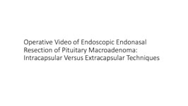 |
Pituitary Surgery | Jonathan Forbes | Operative video of endoscopic endonasal resection of pituitary macroadenoma. Describes intracapsular versus extracapsular techniques. | Pituitary Surgery |
| 86 |
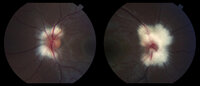 |
Peripapillary Myelinated Nerve Fibers | John J. Chen, MD, PhD | Fundus photographs of a 19-year old female with prominent peripapillary myelinated nerve fibers in both eyes that was incidentally found on routine eye examination. | Myelinated Nerve Fibers |
| 87 |
|
Myelinated Nerve Fibers | John J. Chen, MD, PhD | Fundus photographs of a 19-year old female with prominent peripapillary myelinated nerve fibers in both eyes that was incidentally found on routine eye examination. | Myelinated Nerve Fibers |
| 88 |
 |
Myelinated Nerve Fibers | Carmen Chan,RN, PhD, FAAN | Fundus photos from a patient with extensive myelinated nerve fibers. The patient had normal visual functions. | Myelinated Nerve Fibers |
| 89 |
 |
Common Vitreo-Retinal Procedures and Surgeries | Luke B. Lindsell, OD, MD | Brief presentation on Common vitreo-retinal procedures and surgeries. | Vitreo-Retinal Procedures; Surgeries |
| 90 |
 |
Myotonic Dystrophy | Brian Villafuerte, MD, Ezequiel Piccione, MD | Presentation covering an overview of myotonic dystrophy. | Myotonic Dystrophy |
| 91 |
 |
Muscular Dystrophy Classification | Brian Villafuerte, MD, Ezequiel Piccione, MD | Presentation covering an overview of muscular dystrophy classification. | Muscular Dystrophy Classification |
| 92 |
 |
Congenital Hydrocephalus | Mays El-Dairi, MD | Presentation covering an overview of congenital hydrocephalus. | Congenital Hydrocephalus |
| 93 |
 |
Fibrous Dysplasia | Mays El-Dairi, MD | Presentation covering an overview of fibrous dysplasia. | Fibrous Dysplasia |
| 94 |
 |
Optic Neuropathy: A Recipe for Blindness | Karim Kozhaya, MD; Alaa Bou Ghannam, MD; Alfredo Sadun, MD, PhD | An epidemic of blindness and peripheral neuropathy struck Cuba in the early 90s. By the end of 1993, 7% of the population was affected. Most patients were men and presented with sub-acute, painless, bilateral loss of vision. The etiology of the disease pondered local and international scientists, es... | Cuban Epidemic Optic Neuropathy; Leber's Hereditary Optic Neuropathy; Mitochondrial Insufficiency; Nutritional Optic Neuropathy; Pale Optic Nerve |
| 95 |
 |
Heavy Eye Syndrome | Meagan D. Seay, DO; Bradley J. Katz, MD | A brief overview of heavy eye syndrome. | Heavy Eye Syndrome |
| 96 |
 |
Brown Syndrome | Meagan Seay, DO | A brief overview of Brown Syndrome. | Brown Syndrome |
| 97 |
 |
Intraocular Pressure (IOP) Measurement: A Simple Guide | Nandini Singh; Kirsty Sumerville Mcalester; Alicia Yap; Anne Lee | This video demonstrates the technique for measuring intraocular pressure (IOP) and the use of the tonopen. | Intraocular Pressure (IOP); Tonometry |
1 - 200 of 97